Volume 29, Number 11—November 2023
Synopsis
Outbreak of Pandoraea commovens Infections among Non–Cystic Fibrosis Intensive Care Patients, Germany, 2019–2021
Cite This Article
Citation for Media
Abstract
Pandoraea spp. are gram-negative, nonfermenting rods mainly known to infect patients with cystic fibrosis (CF). Outbreaks have been reported from several CF centers. We report a Pandoraea spp. outbreak comprising 24 non-CF patients at a large university hospital and a neighboring heart center in Germany during July 2019–December 2021. Common features in the patients were critical illness, invasive ventilation, antimicrobial pretreatment, and preceding surgery. Complicated and relapsing clinical courses were observed in cases with intraabdominal infections but not those with lower respiratory tract infections. Genomic analysis of 15 isolates identified Pandoraea commovens as the genetically most similar species and confirmed the clonality of the outbreak strain, designated P. commovens strain LB-19-202-79. The strain exhibited resistance to most antimicrobial drugs except ampicillin/sulbactam, imipenem, and trimethoprim/sulfamethoxazole. Our findings suggest Pandoraea spp. can spread among non-CF patients and underscore that clinicians and microbiologists should be vigilant in detecting and assessing unusual pathogens.
The widespread use of matrix-associated laser desorption/ionization time-of-flight (MALDI-TOF) mass spectrometry, 16s rRNA sequencing, and whole-genome sequencing (WGS) have improved diagnostic accuracy. However, in using those methods, microbiologists and clinicians can be confronted with uncommon gram-negative, nonfermenting bacteria. Those microorganisms originate from the environment, and their pathogenic potential is often unclear. When they are cultured from a clinical specimen, determining whether such a finding represents a true pathogen or a contaminant can be difficult.
One example is the genus Pandoraea, which was described in 2000 when researchers reclassified several Burkholderia- or Ralstonia-like organisms cultured from specimens of patients with cystic fibrosis (CF) (1). Pandoraea spp. are found in soil and water habitats, where the bacteria contribute to soil formation and cycling of elements necessary for plant growth. By 2022, at least 29 species had been identified within the genus, 19 of which were detected in clinical samples, most often from CF patients (2). Those species are P. anapnoica, P. anhela, P. aquatica, P. apista, P. bronchicola, P. capi, P. captiosa, P. cepalis, P. commovens, P. communis, P. faecigallinarum, P. iniqua, P. morbifera, P. nosoerga, P. norimbergensis, P. pneumonica, P. pnomenusa, P. pulmonicola, and P. sputorum (2).
Pandoraea spp. can trigger inflammatory responses and interleukin 6 and 8 elevation in cultures of lung epithelial cells and bacteria from some isolates are capable of crossing lung epithelial cell monolayers (3,4). In an in vivo model for killing Galleria mellonella larvae, virulence of some Pandoraea strains was comparable to that of Burkholderia cenocepacia (3,4). Various other virulence and resistance factors found in other pathogens also can be found in Pandoraea spp. (5,6).
Knowledge on the clinical significance of Pandoraea spp. is based on case reports and case series. Pandoraea spp. can chronically colonize lungs of CF patients and evolve over time by sequential mutations, leading to an adaptation to the CF host niche (7–10). Worsening lung function in CF patients has been linked to Pandoraea spp. colonization, but because CF patients often carry multiple other relevant pathogens, causality between Pandoraea spp. colonization and clinical deterioration is not always clear (9,11,12). The potential of Pandoraea spp. to cause acute illnesses has been exemplified by bloodstream and other life-threatening infections in CF patients and patients who received solid organ transplantation (7,13–15).
Single cases of Pandoraea spp. infections in patient populations other than those with CF or solid organ transplantation have been documented. Cases have occurred among persons without apparent immunodeficiency, causing illnesses such as nosocomial pneumonia, including infections associated with COVID-19, as well as localized hemodialysis catheter infections, prosthetic valve endocarditis, and skull base osteomyelitis (16–22). Nosocomial acquisition and antimicrobial pretreatment seem to be common features among affected patients (16–22).
Pandoraea outbreaks have been documented in CF centers in Denmark and France, each comprising 6 patients (9,12). One in-depth analysis described a large P. apista cluster affecting 18 CF patients serviced at the pediatric and adult CF centers in a city in Scotland and 1 other patient from south England (6). We report a Pandoraea spp. outbreak during July 2019–December 2021 at a large university hospital and the directly neighboring heart center in Berlin, Germany, involving 24 non-CF patients colonized or infected with a novel P. commovens strain.
Patient Data
We retrospectively extracted patient data from hospital records. Data included length of hospital stay, time to isolate Pandoraea spp., antimicrobial drug treatment, intensive care unit (ICU) admission, renal dialysis, solid organ transplantation, underlying conditions exemplified by the Charlson Comorbidity Index (CCI) scores, and patient outcome. We classified patients as either colonized or infected according to the judgment of 2 infectious disease consultants. Detection of Pandoraea spp. from otherwise sterile sites, such as blood, or from intraabdominal specimens was considered as infection. Culture from nonsterile sites (e.g., respiratory samples) was considered colonization if further assessment of antibiotic prescriptions, physicians’ notes, laboratory values, and radiology and pathology findings did not reveal evidence of infection. For the diagnosis of pneumonia, >1 of the following criteria had to be met: new or progressive infiltrate, new or worsening respiratory signs and symptoms, or rising inflammatory markers and assessment of pneumonia by the treating physician. For difficult cases, 2 clinicians discussed and then agreed on a classification for each case (Appendix).
Microbiology
All microbiological investigations were performed by Labor Berlin–Charité Vivantes GmbH in accordance with German Quality Standards for the Microbiological Diagnosis of Infectious Diseases (https://www.dghm.org). To detect aerobic bacteria, we plated clinical specimens on conventional solid media, then incubated in ambient air and 5% CO2 enriched atmosphere at 37°C. We read plates after 24 h and 48 h incubation. We tested suspicious gram-negative microorganisms on oxidase and catalase activity and identified microorganisms by using the VITEK 2 System (bioMérieux, https://www.biomerieux.com), VITEK MALDI-TOF mass spectrometry (bioMérieux), or both. Per our clinical routine, we performed antimicrobial susceptibility testing of Pandoraea spp. by using the VITEK 2 AST GN-233 card, GN-248 card, or both. In addition, we subjected several isolates to further genomic analyses. For those isolates, we performed broth microdilution by using MICRONAUT-S test plates (MERLIN Diagnostika GmbH, https://www.merlin-diagnostika.de), and tested the following antimicrobial agents: piperacillin, piperacillin/tazobactam, temocillin, ceftazidime, cefepime, ceftolozane/tazobactam, ceftazidime/avibactam, meropenem, imipenem, ertapenem, aztreonam, aztreonam/avibactam, ciprofloxacin, levofloxacin, gentamicin, tobramycin, amikacin, trimethoprim/sulfamethoxazole (TMP/SMX), fosfomycin, colistin, minocycline, and tigecycline. We used MIC strips (Liofilchem, https://www.liofilchem.com) to solve discrepancies or to test antimicrobial agents that failed or were not included in VITEK 2 or MICRONAUT-S panels. Were interpreted results according to non–species-related pharmacokinetic/pharmacodynamic (PK/PD) breakpoints published by the European Committee on Antimicrobial Susceptibility Testing (23).
We also conducted environmental investigations for a point source of P. commovens. For environmental investigations, we probed respiratory tubes, nebulizers, suction catheters, washing gloves, toothbrushes, nutrition solutions, inhalation solutions, oral medications such as painkillers in solution, eye and nasal ointments, and eye drops.
Genomic Sequencing and Analysis
For exact species identification and determination of clonality, we subjected 15 clinical outbreak isolates to WGS, by using either the Nextera Flex (Illumina, https://www.illumina.com) or QIASeq FX (QIAGEN, https://www.qiagen.com) library preparation kits, according to the manufacturers’ protocols. In brief, we extracted and enzymatically fragmented 10–100 ng of DNA by using DNeasy PowerSoil Pro Kit (QIAGEN). We added indexed adapters and amplified libraries in limited-cycle PCRs. After clean-up, we quantified, normalized, and pooled sequence-ready libraries before sequencing by using 2× 250 cycles paired-end sequencing on a MiSeq (Illumina).
To close the genome, we performed nanopore sequencing on genomic DNA from isolate LB-19-202-79 from our outbreak on GridION (Oxford Nanopore Technologies, https://nanoporetech.com) using an R9.4 flow cell (Oxford Nanopore). We prepared the sequencing library by using the SQK-LSK109 Ligation Sequencing Kit (Oxford Nanopore), according to the manufacturer’s protocol. We performed basecalling by using Guppy version 5.0.11 (Oxford Nanopore) on the SUP accuracy setting in GridION.
After adapter-trimming the Illumina sequencing reads by using fastp version 0.20.0 (24), we performed de novo genome assemblies by using SPAdes assembler version 3.15.5 (25). For the assembly of nanopore sequencing reads, we tried several protocols using ont-assembly-snake version 1.0 (P. Menzel, unpub. data, https://doi.org/10.20944/preprints202208.0191.v1) and eventually chose the protocol that showed the least differences with P. commovens strain LMG 31010 (National Center for Biotechnology Information [NCBI] RefSeq accession no. GCF_902459615.1). For that protocol, we used Filtong (https://github.com/rrwick/Filtlong) to quality-filter nanopore reads that passed the basecalling quality filter to the top 500 megabases and assembled reads using Flye version 2.9 (26). Then, we polished the initial assembly with the ONT reads by using Racon version 1.4.20 (27) and Medaka version 1.4.3 (https://github.com/nanoporetech/medaka) and polished the Illumina reads by using Polypolish version 0.5.0 (28). We rotated the final assembly to start at the dnaA gene.
We screened the genome assembly of LB-19-202-79 against all available Pandoraea spp. assemblies in the NCBI RefSeq database (29) as of July 21, 2022, by using mash-screen version 2.3 (30). We calculated average nucleotide identity (ANI) between genome assemblies by using FastANI version 1.33 (31). We used andi version 0.12 (32) to calculate pairwise genetic distances between the assembled genomes, from which we constructed a phylogenetic tree comprising the isolate assemblies and the closest Pandoraea spp. by using the neighbor joining method of the ape package version 5.6 (33).
We uploaded the genome assembly to GenBank for annotation by using the NCBI Prokaryotic Genome Annotation Pipeline (34). The uploaded assembly was then automatically annotated by the Bacterial and Viral Bioinformatics Resource Center (BV-BRC) pipeline (https://www.bv-brc.org).
Ethics, Consent, and Permissions
The study was approved by the internal review board of Charité–Universitätmedizin Berlin (registry no. EA4/145/21). The need for informed consent was waived because the study was retrospective.
Figure 1
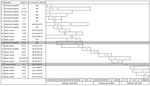
Figure 1. Timeline of an outbreak of Pandoraea commovensamong non–cystic fibrosis intensive care patients, Germany, 2019–2021. The cases clustered at Charité Campus Virchow Klinikum (university hospital) and Deutsches Herzzentrum Berlin...
During July 2019–December 2021, we registered phenotypically identical Pandoraea spp. isolates in specimens from 24 patients, which is 8 times the number of all Pandoraea spp. detected at our laboratory in the 3 previous years (2016–2018). The cases clustered at Charité Campus Virchow Klinikum (CVK) and Deutsches Herzzentrum Berlin (DHZB), 2 neighboring institutions that are on the same grounds; staff and patients regularly move between the 2 institutions. Thirteen patients were treated at DHZB and 9 were treated at CVK. One other patient was treated at Charité Campus Benjamin Franklin and 1 at Unfallkrankenhaus Berlin, a major trauma center; both of those institutions are in different districts of the city. A total of 7 ICUs, 3 at DHZB and 4 at Charité CVK, and 3 regular wards were affected by the outbreak. Cases were first observed at Charité CVK, then the outbreak shifted after a patient was transferred to 2 ICUs at DHZB. Since late August 2019, nearly all isolates have been recovered on those 2 ICUs (Figure 1). Environmental investigations performed at those ICUs in September 2019 did not reveal any point source.
Patient Information and Outcomes
Among the 24 patients whose cultures grew Pandoraea spp., the median age was 67 (range 45–81) years; 50% were male and 50% female. Clinical data were available on 23 patients. Median time from admission to Pandoraea spp. detection was 22 days; 22 (96%) of the 23 patients were treated in ICUs. Patients had numerous underlying conditions, and the median CCI was 6 (range 0–13). All patients had received antimicrobial drug treatment during their hospital stays before Pandoraea spp. detection, 65% had undergone surgery, and 65% received mechanical ventilation (Table 1).
We considered 12 patients colonized and 10 patients infected. For 2 cases, we were unable to make a classification. All 10 of the infected patients were on mechanical ventilation, compared with only 4 (33%) colonized patients. Among infected patients, 8 had lower respiratory tract infections and 2 had intraabdominal infections. In 5 infected patients, Pandoraea spp. was part of a polymicrobial culture, and in the other 5 infected patients, no other pathogens were detected. Pandoraea spp. infections were treated with imipenem in 8 patients and meropenem in 2 patients. TMP/SMX was administered as stepdown therapy in 1 patient after initial treatment with imipenem. All respiratory tract infections resolved, whereas the 2 patients with intraabdominal infections had complicated clinical courses that involved several surgical interventions and protracted administration of various antimicrobial agents in both cases (Appendix).
Among the 10 infected patients, 4 died during hospitalization. None of those deaths were judged to be directly related to the Pandoraea spp. infections (Table 2).
Microbiology Results
During July 2019–December 2021, Pandoraea spp. was cultured from 43 clinical specimens, including throat swabs, respiratory secretions, bile, ascites, intraabdominal specimens, and wound swabs. Numerous blood cultures were collected from 19 of the 24 patients. However, Pandoraea spp. was only detected in 1 blood culture from a patient with a complicated intraabdominal infection. Pandoraea spp. isolates grew readily after overnight culture on commercial solid media, such as Columbia blood or MacConkey agars. Single colonies appeared pale to grayish, displayed weak oxidase activity, and were catalase negative. Neither a mucoid phenotype nor small colony variants were observed.
Using the VITEK 2 GN ID card, identification was possible only to the genus level. In 4 of 17 tested isolates, a reliable discrimination between Pandoraea spp. and Bordetella hinzii could not be made by VITEK 2. In the remaining 13 isolates, we identified Pandoraea spp. with probabilities ranging from 95% to 99%. All 36 isolates tested by VITEK MALDI-TOF mass spectrometry were identified as P. sputorum with a score of 99.9. We were able to perform WGS to differentiate P. commovens from P. sputorum on isolates from patients 7, 9–11, and 14–24, as described in the next section. Because of the local and temporal relationship, identical colony morphology and antimicrobial susceptibilities, we assumed that the correct species identification was P. commovens in all patients reported in this outbreak and that P. sputorum was a misidentification resulting from limitations in the VITEK mass spectrometry database.
We performed susceptibility testing on 35 isolates. For susceptibility testing methods, agreement between VITEK 2 and MICRONAUT-S broth microdilution plates was good for most tested antimicrobial agents. According to EUCAST PK/PD breakpoints, MICs obtained for ampicillin/sulbactam and imipenem were consistently found to be susceptible at standard dosing levels. MICs for TMP/SMX were <20 mg/L in all tested isolates. Cephalosporins including ceftazidime/avibactam, ceftolozane/tazobactam, and cefiderocol tested resistant, as did fluoroquinolones, aminoglycosides, tetracyclines, and colistin (Table 3).
Discrepancies between VITEK 2 and microdilution were apparent for piperacillin and piperacillin/tazobactam; higher MICs were detected using VITEK 2 (range 16 to >128 mg/L for both) than with microdilution (range <4 to 32 mg/L for piperacillin and <1 to 2 mg/L for piperacillin-tazobactam). MICs for meropenem and ertapenem were also higher in VITEK 2 (range 4–8 mg/L and 1 to >8 mg/L, respectively) than in microdilution (range 1–4 mg/L and <0.5 to 1 mg/L, respectively).
Genomic Characterization and Phylogeny
Figure 2
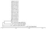
Figure 2. Phylogenetic tree of isolates from an outbreak of Pandoraea commovens among non–cystic fibrosis intensive care patients, Germany, 2019–2021. Genome assemblies from 15 isolates (labeled LB) compared with ...
We screened the genome assemblies from 15 isolates from our outbreak against genomes in RefSeq. We found the genome assembly GCF_902459615.1 of P. commovens strain LMG 31010 to be the most similar, then P. sputorum strain ATCCBAA64, and P. oxalativorans strain DSM 23570. The average genome-wide nucleotide identity between isolate LB-19-202-79 and GCF_902459615.1 was 99.5% and identity between LB-19-202-79 and P. sputorum GCF_900187205.1 was 94.1%. Thus, we designated our strain as P. commovens strain LB-19-202-79. The phylogenetic tree derived from the pairwise phylogenetic distances showed that all our isolates are closely related to each other and distinct from P. commovens strain LMG 31010 and other Pandoraea spp. (Figure 2). We concluded that the outbreak isolates were from a single origin.
The assembly of isolate LB-19-202-79 (NCBI BioSample no. SAMN30015177) from nanopore sequencing data yielded 1 circular chromosome of 5.9-megabase length (GC content 57%; GenBank accession no. CP102780) and 1 plasmid of 80.7 kb length (GC content 63%, 2 copies; GenBank accession no. CP102779).
A search of the plasmid sequence against the NCBI BLAST nucleotide database (https://blast.ncbi.nlm.nih.gov) revealed several alignments to the plasmid of Burkholderia aenigmatica strain CMCC(B)23010 (GenBank accession no. CP091649.1), totaling ≈22 kb and ≈99.9% sequence identity. That strain is a member of the Burkholderia cepacia complex and was originally isolated from water purified for pharmaceuticals. Apart from that, we only found alignments to transposase genes in Klebsiella pneumoniae plasmids.
The annotation of the LB-19-202-79 chromosome yielded 2 β-lactamase family proteins that are also found in the P. commovens strain LMG 31010 genome assembly and have >99% amino acid identity. The β-lactamase with locus tag NTU39_20675 was identified as an oxacillinase (OXA) 62 family carbapenem-hydrolyzing class D enzyme and now is denoted as allele blaOXA-1149 in the NCBI Reference Gene Catalog. The β-lactamase with locus tag NTU39_00730 was identified as a class C β-lactamase. We analyzed the complete resistome comprising all genes associated with antimicrobial resistance as determined by the BV-BRC genome annotation (Appendix Table). The complete genome annotation is available at BV-BRC (accession no. 2508289.5; https://www.bv-brc.org/view/Genome/2508289.5).
We describe a large Pandoraea spp. outbreak comprising 24 non-CF patients. In contrast to earlier outbreaks that took place uniformly among CF patients (6,9,12), none of the patients in this outbreak had CF. However, all but 1 patient were treated in an ICU immediately before or during the time when P. commovens was isolated. All patients had received antimicrobial drugs before P. commovens isolation, and all likely acquired the pathogen in the hospital. Most of the patients had undergone surgery or were on mechanical ventilation, and their overall CCI was high (median 6, range 0–13). Those observations align with earlier case reports on Pandoraea spp. infections among non-CF patients (16–21).
As described by others (35), we experienced difficulties in correctly identifying the species of the outbreak strain. In several cases, P. commovens was misidentified as B. hinzii by biochemical means. Because P. commovens was not identified as a separate species before November 2019 (2), VITEK MALDI-TOF mass spectrometry analysis misidentified the outbreak strain as P. sputorum.
Pandoraea spp. can harbor multiple antimicrobial resistance and biodegradation genes, enabling the pathogen to persist in the hospital environment (6). Our resistome analyses and the course of this outbreak that lasted for 2.5 years suggest that P. commovens LB-19-202-79 is equipped with such an armament.
Pandoraea spp. exhibit resistance to most antimicrobial agents, including penicillins, cephalosporins, fluoroquinolones, aminoglycosides, and colistin, but frequently are susceptible to imipenem and TMP/SMX (14,36,37). Resistance is mediated by different efflux pumps and β-lactamases with a carbapenem-resistant phenotype observed in isolates carrying OXA-62 or a homologue to OXA-153, both carbapenem-hydrolyzing oxacillinases. OXA-62 hydrolyzes meropenem more efficiently than imipenem, but expanded-spectrum cephalosporins are only poor substrates (5,6,38). Among the β-lactamase family proteins detected in our strain, one was identified as an OXA-62 family carbapenem-hydrolyzing class D β-lactamase, now denoted as OXA-1149. However, all our P. commovens isolates were susceptible to imipenem but showed elevated MICs (1–8 mg/L) for meropenem, corresponding to susceptible to increased exposure according to EUCAST PK/PD non–species related breakpoints. The high-level resistance of P. commovens to all cephalosporins might at least in part be mediated by the expression of a class C β-lactamase (39). Although all 8 patients with respiratory tract infections recovered with a single course of antimicrobial drugs, the 2 patients with P. commovens intraabdominal infections had relapsing courses of disease. Pandoraea spp. are environmental bacteria and can thrive in wet settings, such as an abdominal area undergoing multiple surgeries. Under such circumstances, armed with a class D carbapenemase, P. commovens LB-19-202-79 might withstand even prolonged targeted antimicrobial treatment, as noted in patient 7 (Appendix).
The assessment of the clinical significance of detection of P. commovens in the patients in this outbreak was not always straightforward, especially in cases where Pandoraea spp. was cultured from respiratory secretions. Some cases could easily be classified as colonization, such as when only throat swab samples were positive and patients had no other signs and symptoms of infection. However, P. commovens was part of polymicrobial cultures in some patients, and its role in those cases was difficult to estimate. P. commovens was the only relevant pathogen that could be detected in 5 of 8 patients with nosocomial pneumonia; however, it was only detected in low to intermediate quantities. Those cases were classified as infections because patients had nosocomial pneumonia, which cleared after administration of targeted antimicrobial drug treatment for P. commovens. The pathogenicity of P. commovens in the 2 patients with complicated intraabdominal infections seemed evident. P. commovens was the predominant pathogen and was repeatedly isolated from different materials, even from blood culture in 1 case.
The first limitation of this study is that we were not able to sequence all Pandoraea spp. isolates from the outbreak. Some uncertainty remains about whether all isolates belonged to the outbreak strain P. commovens LB-19-202-79. However, given the identical phenotypic features and the temporal and spatial relationship, we assume the same strain was responsible for all cases. Second, we were not able to find an environmental source. Third, detection of Pandoraea spp. could not easily be classified as colonization or infection in several patients; however, that is a well-known dilemma when low-virulent pathogens are cultured from nonsterile sites, such as the respiratory tract.
In conclusion, our sequence analysis highlights the advantage of bacterial WGS for exact species identification and typing of outbreak isolates. On the basis of these findings, we conclude that Pandoraea spp. are not only capable of spreading among CF patients, as described before, but also to non-CF patients. The bacteria can also cause outbreaks on ICUs, in particular affecting patients with a history of intensive antimicrobial pretreatment, multiple abdominal surgeries, and mechanical ventilation. This outbreak report underscores the critical role of vigilance among both clinicians and microbiologists when unusual pathogens occur and the need for access to modern molecular sequencing techniques in hospital laboratories.
Dr. Tassilo Kruis is an internist and medical microbiologist at Labor Berlin Charité Vivantes GmbH, Berlin, Germany. His research interests focus on clinical infectious diseases.
Acknowledgment
All sequencing data are available from the NCBI under BioProject no. PRJNA849608.
References
- Coenye T, Falsen E, Hoste B, Ohlén M, Goris J, Govan JR, et al. Description of Pandoraea gen. nov. with Pandoraea apista sp. nov., Pandoraea pulmonicola sp. nov., Pandoraea pnomenusa sp. nov., Pandoraea sputorum sp. nov. and Pandoraea norimbergensis comb. nov. Int J Syst Evol Microbiol. 2000;50:887–99. DOIPubMedGoogle Scholar
- Peeters C, De Canck E, Cnockaert M, De Brandt E, Snauwaert C, Verheyde B, et al. Comparative genomics of Pandoraea, a genus enriched in xenobiotic biodegradation and metabolism. Front Microbiol. 2019;10:2556. DOIPubMedGoogle Scholar
- Caraher E, Collins J, Herbert G, Murphy PG, Gallagher CG, Crowe MJ, et al. Evaluation of in vitro virulence characteristics of the genus Pandoraea in lung epithelial cells. J Med Microbiol. 2008;57:15–20. DOIPubMedGoogle Scholar
- Costello A, Herbert G, Fabunmi L, Schaffer K, Kavanagh KA, Caraher EM, et al. Virulence of an emerging respiratory pathogen, genus Pandoraea, in vivo and its interactions with lung epithelial cells. J Med Microbiol. 2011;60:289–99. DOIPubMedGoogle Scholar
- Lim Y-L, Ee R, Yong D, Yu C-Y, Ang G-Y, Tee K-K, et al. Complete genome sequence analysis of Pandoraea pnomenusa type strain DSM 16536(T) isolated from a cystic fibrosis patient. Front Microbiol. 2016;7:109. DOIPubMedGoogle Scholar
- Kenna DTD, Coward A, Perry C, Pike R, Schaefer U, Turton J, et al. Investigation of a Pandoraea apista cluster common to adult and paediatric cystic fibrosis patients attending two hospitals in the same city. J Med Microbiol. 2019;68:1081–95. DOIPubMedGoogle Scholar
- Pimentel JD, MacLeod C. Misidentification of Pandoraea sputorum isolated from sputum of a patient with cystic fibrosis and review of Pandoraea species infections in transplant patients. J Clin Microbiol. 2008;46:3165–8. DOIPubMedGoogle Scholar
- Fernández-Olmos A, Morosini MI, Lamas A, García-Castillo M, García-García L, Cantón R, et al. Clinical and microbiological features of a cystic fibrosis patient chronically colonized with Pandoraea sputorum identified by combining 16S rRNA sequencing and matrix-assisted laser desorption ionization-time of flight mass spectrometry. J Clin Microbiol. 2012;50:1096–8. DOIPubMedGoogle Scholar
- Degand N, Lotte R, Decondé Le Butor C, Segonds C, Thouverez M, Ferroni A, et al. Epidemic spread of Pandoraea pulmonicola in a cystic fibrosis center. BMC Infect Dis. 2015;15:583. DOIPubMedGoogle Scholar
- Greninger AL, Streithorst J, Golden JA, Chiu CY, Miller S. Complete genome sequence of sequential Pandoraea apista isolates from the same cystic fibrosis patient supports a model of chronic colonization with in vivo strain evolution over time. Diagn Microbiol Infect Dis. 2017;87:1–6. DOIPubMedGoogle Scholar
- Pugès M, Debelleix S, Fayon M, Mégraud F, Lehours P. Persistent infection because of Pandoraea sputorum in a young cystic fibrosis patient resistant to antimicrobial treatment. Pediatr Infect Dis J. 2015;34:1135–7. DOIPubMedGoogle Scholar
- Jørgensen IM, Johansen HK, Frederiksen B, Pressler T, Hansen A, Vandamme P, et al. Epidemic spread of Pandoraea apista, a new pathogen causing severe lung disease in cystic fibrosis patients. Pediatr Pulmonol. 2003;36:439–46. DOIPubMedGoogle Scholar
- Johnson LN, Han J-Y, Moskowitz SM, Burns JL, Qin X, Englund JA. Pandoraea bacteremia in a cystic fibrosis patient with associated systemic illness. Pediatr Infect Dis J. 2004;23:881–2. DOIPubMedGoogle Scholar
- Stryjewski ME, LiPuma JJ, Messier RH Jr, Reller LB, Alexander BD. Sepsis, multiple organ failure, and death due to Pandoraea pnomenusa infection after lung transplantation. J Clin Microbiol. 2003;41:2255–7. DOIPubMedGoogle Scholar
- Xiao X, Tian H, Cheng X, Li G, Zhou J, Peng Z, et al. Pandoraea sputorum bacteremia in a patient who had undergone allogeneic liver transplantation plus immunosuppressive therapy: a case report. Infect Drug Resist. 2019;12:3359–64. DOIPubMedGoogle Scholar
- Lin C, Luo N, Xu Q, Zhang J, Cai M, Zheng G, et al. Pneumonia due to Pandoraea Apista after evacuation of traumatic intracranial hematomas:a case report and literature review. BMC Infect Dis. 2019;19:869. DOIPubMedGoogle Scholar
- Monzón T, Valga F, Reichert J, López C. Hemodialysis catheter colonized by Pandoraea spotorum. [Engl Ed]. Nefrologia (Engl Ed). 2018;38:662–4. DOIPubMedGoogle Scholar
- Bodendoerfer E, Personnic N, Mestres CA, Wilhelm MJ, Meyer L, Hasse B. Possible prosthetic valve endocarditis by Pandoraea pnomenusa and specific virulence mechanisms. Infect Drug Resist. 2021;14:1319–24. DOIPubMedGoogle Scholar
- Gawalkar AA, Kasinadhuni G, Kanaujia R, Rajan P, Vijay J, Revaiah PC, et al. Prosthetic aortic valve dehiscence following infective endocarditis by a rare bacterium - Pandoraea pnomenusa. J Cardiol Cases. 2020;24:27–9. DOIPubMedGoogle Scholar
- Patil NR, Tripathi M, Charaya K, Angrup A, Ahuja C, Mohindra S. Skull base osteomyelitis by Pandoraea apista: An unusual pathogen at unusual location - A case report. Surg Neurol Int. 2021;12:447. DOIPubMedGoogle Scholar
- Singh S, Sahu C, Patel SS, Garg A, Ghoshal U. Pandoraea apista bacteremia in a COVID-positive man: a rare coinfection case report from north India. J Lab Physicians. 2021;13:192–4. DOIPubMedGoogle Scholar
- Dlewati MM, Aung PP, Park K, Rodriguez JA, Poon KK. Meropenem-resistant Pandoraea pneumonia in a critically ill patient with COVID-19. Cureus. 2021;13:
e19498 . DOIPubMedGoogle Scholar - European Committee on Antimicrobial Susceptibility Testing. EUCAST Clinical Breakpoint Tables version 11.0, 2021 [cited 2021 Dec 31]. https://www.eucast.org/fileadmin/src/media/PDFs/EUCAST_files/Breakpoint_tables/v_11.0_Breakpoint_Tables.pdf
- Chen S, Zhou Y, Chen Y, Gu J. fastp: an ultra-fast all-in-one FASTQ preprocessor. Bioinformatics. 2018;34:i884–90. DOIPubMedGoogle Scholar
- Prjibelski A, Antipov D, Meleshko D, Lapidus A, Korobeynikov A. Using SPAdes de novo assembler. Curr Protoc Bioinformatics. 2020;70:
e102 . DOIPubMedGoogle Scholar - Kolmogorov M, Yuan J, Lin Y, Pevzner PA. Assembly of long, error-prone reads using repeat graphs. Nat Biotechnol. 2019;37:540–6. DOIPubMedGoogle Scholar
- Vaser R, Sović I, Nagarajan N, Šikić M. Fast and accurate de novo genome assembly from long uncorrected reads. Genome Res. 2017;27:737–46. DOIPubMedGoogle Scholar
- Wick RR, Holt KE. Polypolish: Short-read polishing of long-read bacterial genome assemblies. PLOS Comput Biol. 2022;18:
e1009802 . DOIPubMedGoogle Scholar - O’Leary NA, Wright MW, Brister JR, Ciufo S, Haddad D, McVeigh R, et al. Reference sequence (RefSeq) database at NCBI: current status, taxonomic expansion, and functional annotation. Nucleic Acids Res. 2016;44(D1):D733–45. DOIPubMedGoogle Scholar
- Ondov BD, Starrett GJ, Sappington A, Kostic A, Koren S, Buck CB, et al. Mash Screen: high-throughput sequence containment estimation for genome discovery. Genome Biol. 2019;20:232. DOIPubMedGoogle Scholar
- Jain C, Rodriguez-R LM, Phillippy AM, Konstantinidis KT, Aluru S. High throughput ANI analysis of 90K prokaryotic genomes reveals clear species boundaries. Nat Commun. 2018;9:5114. DOIPubMedGoogle Scholar
- Haubold B, Klötzl F, Pfaffelhuber P. andi: fast and accurate estimation of evolutionary distances between closely related genomes. Bioinformatics. 2015;31:1169–75. DOIPubMedGoogle Scholar
- Paradis E, Schliep K. ape 5.0: an environment for modern phylogenetics and evolutionary analyses in R. Bioinformatics. 2019;35:526–8. DOIPubMedGoogle Scholar
- Li W, O’Neill KR, Haft DH, DiCuccio M, Chetvernin V, Badretdin A, et al. RefSeq: expanding the Prokaryotic Genome Annotation Pipeline reach with protein family model curation. Nucleic Acids Res. 2021;49(D1):D1020–8. DOIPubMedGoogle Scholar
- Martina PF, Martínez M, Frada G, Alvarez F, Leguizamón L, Prieto C, et al. First time identification of Pandoraea sputorum from a patient with cystic fibrosis in Argentina: a case report. BMC Pulm Med. 2017;17:33. DOIPubMedGoogle Scholar
- Daneshvar MI, Hollis DG, Steigerwalt AG, Whitney AM, Spangler L, Douglas MP, et al. Assignment of CDC weak oxidizer group 2 (WO-2) to the genus Pandoraea and characterization of three new Pandoraea genomospecies. J Clin Microbiol. 2001;39:1819–26. DOIPubMedGoogle Scholar
- Fernández-Olmos A, Morosini MI, Lamas A, García-Castillo M, García-García L, Cantón R, et al. Clinical and microbiological features of a cystic fibrosis patient chronically colonized with Pandoraea sputorum identified by combining 16S rRNA sequencing and matrix-assisted laser desorption ionization-time of flight mass spectrometry. J Clin Microbiol. 2012;50:1096–8. DOIPubMedGoogle Scholar
- Schneider I, Queenan AM, Bauernfeind A. Novel carbapenem-hydrolyzing oxacillinase OXA-62 from Pandoraea pnomenusa. Antimicrob Agents Chemother. 2006;50:1330–5. DOIPubMedGoogle Scholar
- Philippon A, Arlet G, Labia R, Iorga BI. Class C β-lactamases: molecular characteristics. Clin Microbiol Rev. 2022;35:
e0015021 . DOIPubMedGoogle Scholar
Figures
Tables
Cite This ArticleOriginal Publication Date: October 17, 2023
Table of Contents – Volume 29, Number 11—November 2023
| EID Search Options |
|---|
|
|
|
|
|
|
Please use the form below to submit correspondence to the authors or contact them at the following address:
Tassilo Kruis, Charité Vivantes GmbH, Sylter Str. 2, Berlin 13353, Germany
Top