Volume 29, Number 4—April 2023
Research Letter
Highly Pathogenic Avian Influenza A(H5N1) Virus in a Harbor Porpoise, Sweden
Elina Thorsson, Siamak Zohari, Anna Roos, Fereshteh Banihashem, Caroline Bröjer, and Aleksija Neimanis
Figure 1
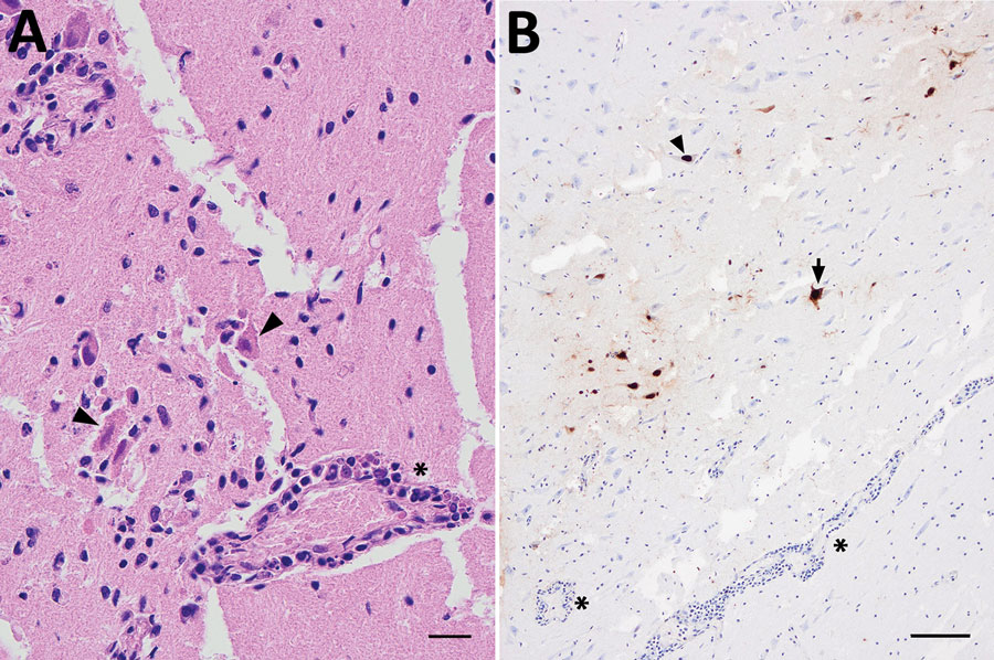
Figure 1. Microscopic analyses of tissue samples from a harbor porpoise (Phocoena phocoena) infected with highly pathogenic avian influenza virus H5N1 clade 2.3.4.4b, Sweden. A) Brain tissue showing neuronal necrosis (arrowheads) and perivascular lymphoplasmacytic cuffing of vessels and vasculitis (asterisk). Scale bar represents 20 µm. B) Immunohistochemical labeling of influenza A nucleoprotein in neuronal nuclei (arrowhead) and cytoplasm (arrow), as well as glial cells. Perivascular cuffing (asterisks) is seen in close association to influenza A immunolabeling. Scale bar represents 100 µm.
Page created: January 24, 2023
Page updated: March 21, 2023
Page reviewed: March 21, 2023
The conclusions, findings, and opinions expressed by authors contributing to this journal do not necessarily reflect the official position of the U.S. Department of Health and Human Services, the Public Health Service, the Centers for Disease Control and Prevention, or the authors' affiliated institutions. Use of trade names is for identification only and does not imply endorsement by any of the groups named above.