Skip directly to site content Skip directly to page options Skip directly to A-Z link Skip directly to A-Z link Skip directly to A-Z link
Volume 29, Number 6—June 2023
Dispatch

Detection of Novel Poxvirus from Gray Seal (Halichoerus grypus), Germany

Florian Pfaff, Katharina Kramer, Jacqueline King, Kati Franzke, Tanja Rosenberger, Dirk Höper, Patricia König, Donata Hoffmann, and Martin BeerComments to Author 
Author affiliations: Friedrich-Loeffler-Institute, Greifswald, Germany (F. Pfaff, J. King, K. Franzke, D. Höper, P. König, D. Hoffmann, M. Beer); Landeslabor Schleswig-Holstein, Neumünster, Germany (K. Kramer); Seehundstation Friedrichskoog e.V., Friedrichskoog, Germany (T. Rosenberger)

Main Article

Figure 1

Histopathology and electron microscopy of nodules and lung tissue from a gray seal (Halichoerus grypus) with novel poxvirus, North Sea, Germany. A) Histopathology of nodules shows severe papillary epithelial hyperplasia with infiltration of neutrophils. Scale bar indicates 200 μm. B) Histopathology of ballooning degeneration of epithelial cells. Arrows indicate large eosinophilic intracytoplasmic inclusion bodies. Scale bar indicates 50 μm. C) Histopathology of the lung shows multifocal moderate pneumonia with infiltration of mononuclear cells and neutrophils with proliferation of pneumocytes type II and intra-alveolar histiocytosis, severe atelectasis, and hyperemia. Scale bar indicates 100 μm. D) Negative-contrast electron microscopy of lung tissue. Microscopy revealed poxvirus-like viral particles. Scale bar indicates 300 nm. Inset: closeup of poxvirus-like particles, which had an oval shape ≈250 nm × ≈200 nm and an irregular surface with randomly arranged tubular structures; scale bar indicates 100 nm.

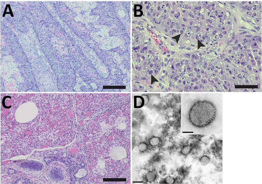
Figure 1. Histopathology and electron microscopy of nodules and lung tissue from a gray seal (Halichoerus grypus) with novel poxvirus, North Sea, Germany. A) Histopathology of nodules shows severe papillary epithelial hyperplasia with infiltration of neutrophils. Scale bar indicates 200 μm. B) Histopathology of ballooning degeneration of epithelial cells. Arrows indicate large eosinophilic intracytoplasmic inclusion bodies. Scale bar indicates 50 μm. C) Histopathology of the lung shows multifocal moderate pneumonia with infiltration of mononuclear cells and neutrophils with proliferation of pneumocytes type II and intra-alveolar histiocytosis, severe atelectasis, and hyperemia. Scale bar indicates 100 μm. D) Negative-contrast electron microscopy of lung tissue. Microscopy revealed poxvirus-like viral particles. Scale bar indicates 300 nm. Inset: closeup of poxvirus-like particles, which had an oval shape ≈250 nm × ≈200 nm and an irregular surface with randomly arranged tubular structures; scale bar indicates 100 nm.

Main Article

Page created: March 29, 2023
Page updated: May 17, 2023
Page reviewed: May 17, 2023
The conclusions, findings, and opinions expressed by authors contributing to this journal do not necessarily reflect the official position of the U.S. Department of Health and Human Services, the Public Health Service, the Centers for Disease Control and Prevention, or the authors' affiliated institutions. Use of trade names is for identification only and does not imply endorsement by any of the groups named above.
file_external