Volume 29, Number 6—June 2023
Dispatch
Ranid Herpesvirus 3 Infection in Common Frog Rana temporaria Tadpoles
Francesco C. Origgi and Annette Taugbøl
Figure 1
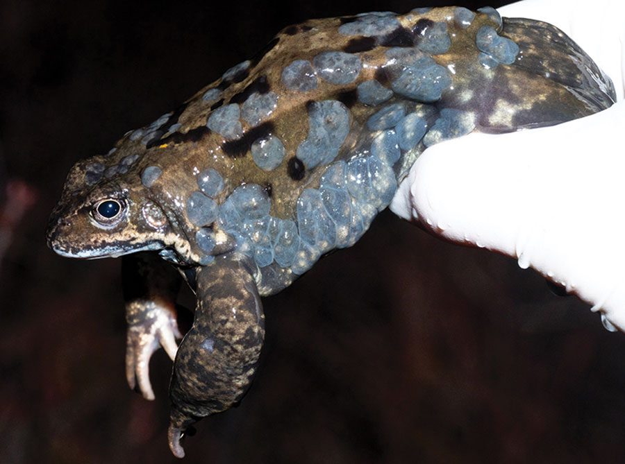
Figure 1. Ranid herpesvirus 3 infection in common frog Rana temporaria tadpoles, Norway. Image shows a large number of multifocal to coalescent, mildly elevated, gray patches (epidermal hyperplasia) extending over most of the integument, particularly clustering along the left flank. Between gray areas is normally pigmented skin. Image copyright © Jeroen van der Kooij.
Page created: April 12, 2023
Page updated: May 17, 2023
Page reviewed: May 17, 2023
The conclusions, findings, and opinions expressed by authors contributing to this journal do not necessarily reflect the official position of the U.S. Department of Health and Human Services, the Public Health Service, the Centers for Disease Control and Prevention, or the authors' affiliated institutions. Use of trade names is for identification only and does not imply endorsement by any of the groups named above.