Tanapox, South Africa, 2022
Monica Birkhead
1
, Wayne Grayson
1, Antoinette Grobbelaar, Veerle Msimang, Naazneen Moolla, Angela Mathee, Lucille Blumberg, Terry Marshall, Daniel Morobadi, Mirjana Popara, and Jacqueline Weyer
Author affiliations: National Institute for Communicable Diseases, Johannesburg, South Africa (M. Birkhead, A. Grobbelaar, V. Msimang, N. Moolla, L. Blumberg, J. Weyer); Ampath Laboratories, Centurion, South Africa (W. Grayson, T. Marshall, D. Morobadi); University of the Witwatersrand, Johannesburg (W. Grayson, J. Weyer); South African Medical Research Council, Cape Town, South Africa (A. Mathee); University of Johannesburg, Johannesburg (A. Mathee); University of Pretoria, Pretoria, South Africa (N. Moolla, L. Blumberg, J. Weyer); Right to Care, Johannesburg (L. Blumberg); University of the Free State, Bloemfontein, South Africa (D. Morobadi); Mediclinic, Sandton, South Africa (M. Popara)
Main Article
Figure 2
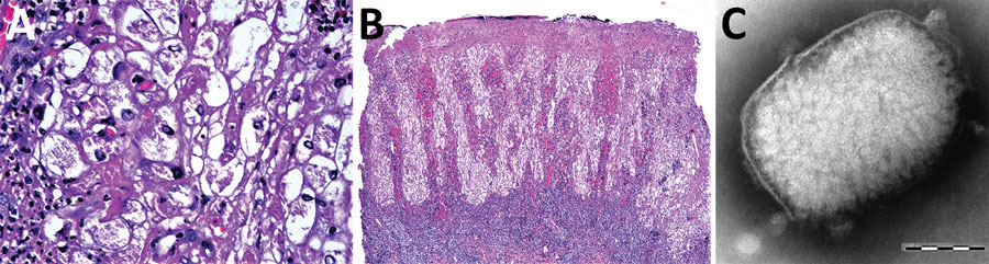
Figure 2. Diagnostic light and electron microscopy of tanapox lesion biopsies from a case-patient, South Africa, 2022. A) High-power photomicrograph of initial skin biopsy, showing prominent vacuolation of epidermal keratinocytes, granular intracytoplasmic inclusions, and intranuclear pseudoinclusions. Hematoxylin and eosin stain; original magnification ×400. B) Low-power photomicrograph of initial skin biopsy, showing a superficially eroded hyperplastic epidermis, with cytoplasmic pallor and a dense underlying superficial dermal lymphoid infiltrate. Hematoxylin and eosin stain; original magnification ×40. C) Negatively stained tanapox virus virion with surface tubules evident beneath the remains of the surrounding membrane. Virion dimensions were 159–327 nm × 186–289 nm. Scale bar indicates 100 nm.
Main Article
Page created: March 28, 2023
Page updated: May 17, 2023
Page reviewed: May 17, 2023
The conclusions, findings, and opinions expressed by authors contributing to this journal do not necessarily reflect the official position of the U.S. Department of Health and Human Services, the Public Health Service, the Centers for Disease Control and Prevention, or the authors' affiliated institutions. Use of trade names is for identification only and does not imply endorsement by any of the groups named above.