Volume 29, Number 6—June 2023
Dispatch
Tanapox, South Africa, 2022
Cite This Article
Citation for Media
Abstract
Tanapox is a rarely diagnosed zoonosis known to be endemic to equatorial Africa. All previously reported human cases were acquired within 10° north or south of the Equator, most recently 19 years ago. We describe a human case of tanapox in South Africa (24° south of the Equator). Expanded surveillance for this pathogen is warranted.
Tanapox is a rarely diagnosed zoonosis endemic to equatorial Africa. Only 4 exported cases from Africa involving either human or nonhuman primates have been reported (Table). Initial human outbreaks, in 1957 and 1962, were recorded from the Tana River Valley of Kenya, and the etiologic agent was subsequently isolated and described as Tanapox virus (TANV) (genus Yatapoxvirus, family Poxviridae) in 1965 (1). Subsequently, the occurrence of tanapox in research laboratory primates imported into the United States (2,3) led to a serologic survey of 12 nonhuman primate species from Kenya, Ethiopia, Cameroon, Côte d’Ivoire, Liberia, and Senegal; seropositivity was detected in all species surveyed in those countries (8). Therefore, nonhuman primates across equatorial Africa were surmised to be the natural reservoirs of TANV and humans incidental hosts (3,8). On the basis of the overlap between human tanapox cases and the geographic ranges of selected nonhuman primates, an ecologic niche model predicted that tanapox could be found from Somalia to Senegal, with the most southerly range above the Tropic of Capricorn (9).
The epidemiology and natural ecology of tanapox is largely unknown, but previous reports indicate that all infected humans are equally affected, regardless of age group and sex. Serologic surveys conducted in Tana River communities indicated 16.3% prevalence in 1971 and 9.2% in 1976 (10). Human-to-human transmission is rare, and although transmission from nonhuman primates to humans by contact or inoculation has been noted under laboratory conditions, natural human infections are more likely to be acquired by mechanical transmission from contaminated mouthparts of hematophagous arthropods (2–4). This vector theory arose because of the synchronicity between tanapox outbreaks and the increased arthropod activity associated with seasonal high temperatures, high rainfall, and flooding in the riparian areas in which surveillance was done (1,3,9). The similarities in the distribution and incidence of TANV and West Nile virus antibodies in serum samples collected in the Tana River Valley in 1971 led to the suggestion that both viruses are transmitted in the same way (i.e., by a culicine mosquito, probably a species of Mansonia) (1,3).
In humans, tanapox typically manifests with 1 or 2 characteristic, nodular skin lesions that are large, raised, umbilicated, and painful and generally ulcerate without becoming pustular (in contrast to lesions observed in most other poxvirus infections) (1,3,7). The lesions may be associated with localized lymphadenopathy, and their gradual development is preceded by a mild, short-lived febrile illness, with possible pruritus and myalgia leading to prostration and headaches (1–3,5,6). Histologically, lesions are restricted to the epithelial layers, with cells containing eosinophilic cytoplasmic inclusions and vacuolated nuclei (1,2,7). TANV virions are poxlike but cannot be distinguished microscopically from the other species in the genus (e.g., Yaba monkey tumor virus) or from Orthopoxvirus virions (3,7). Clinical differential diagnoses have included cutaneous anthrax, other poxvirus infections, sporotrichosis, Mycobacterium marinum infection, spotted fever group rickettsial infections, tropical ulcers, insect bites, and scabies (3,7,9). The disease is self-limiting, with no recorded fatalities (3). We describe a case of tanapox in South Africa, 19 years after the last published report of human tanapox (7).
Figure 1
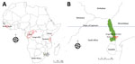
Figure 1. Geographic distribution of recorded human cases of tanapox. A) Locations of previous tanapox cases reported in the literature. Red dots indicate cases acquired locally; red outlines indicate regions of countries...
We obtained written consent from the patient in this study and received ethics clearance from the Faculty of Health Sciences of University of the Witwatersrand, Johannesburg (approval no. M210752). During February 2–6, 2022, a 61-year old woman served as a volunteer in Kruger National Park (KNP), South Africa. She stayed in a tented bush camp along the banks of the Sand River, ≈20 km from the town of Skukuza (24°59′43′′ S, 31°35′34′′E) (Figure 1; Appendix Figure 1). The woman noted large numbers of arthropods (e.g., spiders, insects, and ticks) around the camp site and reported being bitten on several occasions on various body parts (especially on her hands, shoulders, arms, and back). Because of heavy rains, trails were overgrown, so bushwalks resulted in scratches on her arms, and many ticks were found on her clothing. During February 7–9, she continued her visit to KNP as a guest in air-conditioned accommodation and had no further direct contact with vegetation. She reported no direct contact with primates, although vervet monkeys (Chlorocebus pygerythrus) are seen near the camps.
Two days after her return to urban Johannesburg, she experienced pruritus at the base of her thumb on the dorsal side of her right hand and noticed a pale blister forming there, followed 2 days later by another blister on the side of her left hand. Initially, both papules were round and white with erythematous edges (Appendix Figure 2, panel A), but they became dome-shaped, firm, smooth, umbilicated nodules 12–15 mm in size (Appendix Figure 2, panels B, C). A third lesion formed on the woman’s mid-upper back but was perforated through chafing from clothing. About 3 days after the appearance of the first lesion, the woman reported feeling unwell, fatigued, and feverish and had severe headaches. No lymphadenopathy was recorded. The lesions were persistently painful and hypersensitive, but none were cystic or became pustular. Instead, they became ulcerated, open, and dry (Appendix Figure 2, panel D), and all 3 resolved over a period of 6 weeks, leaving slight discoloration. After discovering the third lesion, the woman sought medical attention. Differential diagnoses included allergies, cellulitis, erysipelas, pyoderma gangrenosum, or granulomas caused by foreign bodies or insect bites (e.g., mango fly bites).
Figure 2
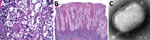
Figure 2. Diagnostic light and electron microscopy of tanapox lesion biopsies from a case-patient, South Africa, 2022. A) High-power photomicrograph of initial skin biopsy, showing prominent vacuolation of epidermal keratinocytes, granular intracytoplasmic...
Histopathologic examination of a lesion biopsy indicated a possible pox infection, given the presence of acidophilic intracytoplasmic inclusion bodies and cellular vacuolation (Figure 2, panel A). The lesion was confined to the epithelial layers with many cells that had vacuolated nuclei, and we observed ballooning cells in the deeper epithelial layer (Figure 2, panel B). Subsequently, we took an additional biopsy for electron microscopy and 2 lesion swab specimens for molecular characterization. After routine processing (Appendix), we observed numerous brick-shaped virions 287 nm x 221 nm with distinct surface tubules and generally an outer membrane layer (capsular form) (Figure 2, panel C). We used 1 dry swab sample for PCR analysis (Appendix), and partial sequence analysis indicated clustering with available TANV sequences (Appendix Figure 3). Attempts at full genomic sequencing and virus isolation (from the second swab specimen) were unsuccessful, possibly because of the limited clinical material available.
Recent surveys of the mosquito distribution in southern Africa have found that 2 culicine genera (Culex and Mansonia) comprise 91% of the mosquito population in the town of Skukuza (11). Weather conditions at the time of the case exposure were conducive to vector replication; recent rainfall was up to 147% higher than the average annual cumulative total (12), and ambient temperatures were 100°F–104°F (38°C–40°C) (13). In terms of virus reservoirs, a limitation of Monroe et al.’s model (9) was that the restricted range of recorded human cases determined the exclusion of many other primates with extensive geographic ranges. Our report extends this range beyond the most southerly predictions of the model, which increases the pool of potential reservoir hosts.
The clinical findings for this reported case were in keeping with previous clinical reports of tanapox, and the diagnosis was supported by histopathology, electron microscopy, and molecular analyses. The importance and continuing relevance of histopathology and microscopy to the diagnosis and investigation of zoonotic disease were clearly illustrated. Given that tanapox is a vectorborne disease, many drivers, including anthropogenic destruction of wildlife habitats, environmental instability, and global climate change, may influence its emergence (14). Improved surveillance, including studies relating to the ecology and epidemiology of TANV in vectors, hosts, and humans, is warranted.
Dr. Birkhead is a medical scientist in the electron microscopy laboratory of the National Institute for Communicable Diseases, a division of the National Health Laboratory Service, in Johannesburg, South Africa. Her primary research interests include the use of ultrastructural information for diagnoses of and research on human infectious diseases.
Acknowledgment
We thank Nosihle Msomi for her support in sequencing and sequencing analysis performed in this study.
References
- Downie AW, Taylor-Robinson CH, Caunt AE, Nelson GS, Manson-Bahr PEC, Matthews TCH. Tanapox: a new disease caused by a pox virus. BMJ. 1971;1:363–8. DOIPubMedGoogle Scholar
- Downie AW, España C. A comparative study of Tanapox and Yaba viruses. J Gen Virol. 1973;19:37–49. DOIPubMedGoogle Scholar
- Jezek Z, Arita I, Szczeniowski M, Paluku KM, Kalisa R, Nakano JH. Human tanapox in Zaire: clinical and epidemiological observations on cases confirmed by laboratory studies. Bull. World Health Organ. 1985;63:1027–35.
- Downie AW, España C. Comparison of Tanapox virus and Yaba-like viruses causing epidemic disease in monkeys. J Hyg (Lond). 1972;70:23–32. DOIPubMedGoogle Scholar
- Croitoru AG, Birge MB, Rudikoff D, Tan MH, Phelps RG. Tanapox virus infection. Skinmed. 2002;1:156–7. DOIPubMedGoogle Scholar
- Stich A, Meyer H, Köhler B, Fleischer K. Tanapox: first report in a European traveller and identification by PCR. Trans R Soc Trop Med Hyg. 2002;96:178–9. DOIPubMedGoogle Scholar
- Dhar AD, Werchniak AE, Li Y, Brennick JB, Goldsmith CS, Kline R, et al. Tanapox infection in a college student. N Engl J Med. 2004;350:361–6. DOIPubMedGoogle Scholar
- Downie AW. Serological evidence of infection with Tana and Yaba pox viruses among several species of monkey. J Hyg (Lond). 1974;72:245–50. DOIPubMedGoogle Scholar
- Monroe BP, Nakazawa YJ, Reynolds MG, Carroll DS. Estimating the geographic distribution of human Tanapox and potential reservoirs using ecological niche modeling. Int J Health Geogr. 2014;13:34. DOIPubMedGoogle Scholar
- Axford JS, Downie AW. Tanapox. A serological survey of the lower Tana River Valley. J Hyg (Lond). 1979;83:273–6. DOIPubMedGoogle Scholar
- Cornel AJ, Lee Y, Almeida APG, Johnson T, Mouatcho J, Venter M, et al. Mosquito community composition in South Africa and some neighboring countries. Parasit Vectors. 2018;11:331. DOIPubMedGoogle Scholar
- SANparks Scientific Services. Data and information resources: Kruger climate and rainfall [cited 2023 Mar 27]. https://www.sanparks.org/scientific-services/wp-content/uploads/2022/01/December.pdf
- Meteoblue. Weather history and climate archive [cited 2023 Mar 27]. https://www.meteoblue.com/en/weather/historyclimate/weatherarchive/skukuza_south-africa_954955
- Carlson CJ, Albery GF, Merow C, Trisos CH, Zipfel CM, Eskew EA, et al. Climate change increases cross-species viral transmission risk. Nature. 2022;607:555–62. DOIPubMedGoogle Scholar
Figures
Table
Cite This ArticleOriginal Publication Date: April 06, 2023
1These first authors contributed equally to this article.
Table of Contents – Volume 29, Number 6—June 2023
| EID Search Options |
|---|
|
|
|
|
|
|
Please use the form below to submit correspondence to the authors or contact them at the following address:
Monica Birkhead, National Institute for Communicable Diseases, Private Bag X4, Sandringham, 2192, South Africa
Top