Volume 29, Number 7—July 2023
Dispatch
Surveillance and Genomic Characterization of Influenza A and D Viruses in Swine, Belgium and the Netherlands, 2019–2021
Cite This Article
Citation for Media
Abstract
During 2019–2021, we isolated 62 swine influenza A viruses in Belgium and the Netherlands. We also detected influenza D in pigs in the Netherlands. The ever-changing diversity of influenza viruses and the identification of influenza D emphasize the need for more virus surveillance.
Three influenza A virus (IAV) subtypes circulate globally in swine: H1N1, H1N2 and H3N2. Within each subtype, multiple hemagglutinin (HA) and neuraminidase (NA) lineages or clades cocirculate. In Europe, there are 4 swine IAV HA lineages: the HA-1A classical swine lineage including influenza A(H1N1)pdm09 virus (pH1N1), HA-1B human seasonal lineage (H1hu), HA-1C Eurasian avian lineage (H1av), and European human-like H3 lineage (1). In addition, there are 4 NA lineages: pH1N1, N1av, A/swine/Scotland/410440/1994-like (N2s), and A/swine/Gent/1/1984-like (N2g). Swine IAV lineages have continued to evolve through exchange of genome segments (reassortment) and mutations in the viral surface proteins (drift); these processes resulted in multiple genotypes with gene segments of swine, avian, and human origin.
A new genus, influenza D virus (IDV), was identified in pigs in Oklahoma in 2011 (2). Two IDV lineages, D/swine/Oklahoma/1334/2011 and D/bovine/Oklahoma/660/2013 (D/660), have been identified in swine and cattle in Europe on the basis of the hemagglutinin-esterase fusion protein (HEF). In this study, we identified the prevailing swine IAVs circulating in Belgium and the Netherlands during 2019–2021 and determined their genotypes by whole-genome characterization. Furthermore, we described the isolation and characterization of an emerging swine IDV in the Netherlands.
We started with 152 samples that had tested IAV-positive by reverse transcription PCR by Animal Healthcare Flanders (Torhout, Belgium) or through random viral and bacterial metagenomics analysis by PathoSense (Lier, Belgium) (3). The samples were submitted to the diagnostic laboratories by the farmer or the veterinarian for an etiologic diagnosis of respiratory signs. Samples were nasal and tracheobronchial swab specimens and lung tissues from pigs in Belgium and the Netherlands, collected during November 2019–December 2021. From those samples, we obtained 62 swine IAVs by virus isolation in MDCK cells. We determined their subtype by hemagglutination inhibition assays with reference antiserum that we obtained by double vaccination with inactivated virus vaccines (Appendix 1 Table) and a multiplex reverse transcription quantitative PCR, specific for the HA and NA (Table 1; Appendix 1 Table) (4). Two of those IAVs originated from pigs in the Netherlands; the other 60 were isolated from pigs in Belgium. Subtyping revealed 34 (54.8%) viruses of the H1N1 subtype, comprising 30 (48.4%) H1avN1av viruses, 3 (4.8%) pH1N1av viruses, and 1 (1.6%) H1huN1av virus. The second most dominant subtype was H1N2 with 27 (43.5%) viruses: 13 (21.0%) H1huN2 viruses, 13 (21.0%) pH1N2 viruses, and 1(1.6%) H1avN2 virus. We isolated 1 (1.6%) H3N2 virus.
We selected 23 of the 62 swine IAV isolates for targeted whole-genome sequencing using nanopore sequencing (Table 2) (5–7). All sequences are available through GenBank (accession nos. OP445672–79, OP445741–812, OP458600–702). Sequencing of HA revealed that the isolates belonged to all 4 lineages reported in Europe: 10 H1av, 8 pH1, 4 H1hu, and 1 H3. For NA, all isolates of the H1N1 subtype had their NA derived from H1av viruses. Within the H1N2 subtype, 2 isolates derived their NA from N2s and 9 from N2g viruses. The 6 internal gene segments were derived from pH1 or H1av viruses or a combination of both. We designated the A/swine/Belgium/Gent-249/2020 genotype, previously reported by Chepkwony et al. (7), as AQ; we identified a genotype of isolate A/swine/Belgium/Gent-234/2020 and designated it as AR. The naming of both isolates is in accordance with the nomenclature system introduced by Watson (8) and updated by Henritzi (9).
Figure 1
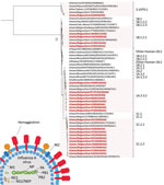
Figure 1. Phylogenetic tree based on the hemagglutinin nucleotide sequences of 23 swine influenza A isolates from pigs in Belgium and the Netherlands, November 2019–December 2021, and 28 swine and human influenza...
Figure 2
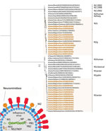
Figure 2. Phylogenetic tree based on neuraminidase nucleotide sequences of 23 swine influenza A isolates from pigs in Belgium and the Netherlands, November 2019–December 2021, and 28 swine and human influenza A...
For both HA and NA sequences, we performed maximum-likelihood phylogenetic analyses (10) (Figures 1, 2). We selected a total of 28 swine and human IAVs as reference viruses, including both historical and contemporary IAVs circulating in pigs as well as humans in Europe and North America; sources isolates were US Department of Agriculture, Worldwide Influenza Centre at Francis Crick Institute, US Centers for Disease Control, the Ploufragan-Plouzané-Niort Laboratory of the French Agency for Food, Environmental and Occupational Health & Safety(, and the Istituto Zooprofilattico Sperimentale delle Venezie. This analysis revealed that 8 of 10 H1av isolates belonged to clade 1C.2.2 and 2 isolates to clade 1C.2.1. In contrast, all 4 H1hu isolates belonged to clade 1B.1.2.1 and all 8 pH1 isolates to clade 1A.3.3.2. The NA genes grouped per lineage and the shorter horizontal branches suggest a slower evolution than the HA gene.
Two tracheobronchial swab specimens from swine farms, originating from Belgium and the Netherlands, tested IDV-positive via metagenomics in 2021 (3). The pigs had demonstrated mild upper respiratory signs. Cattle were present on both farms. The samples were investigated by virus isolation on swine testicle cells. We isolated IDV from the sample from the Netherlands and performed next-generation whole-genome sequencing. We designated the isolate as D/swine/Netherlands/PS-497/2021 (GenBank accession nos. OP474071–77); phylogenetic analyses of the HEF nucleotide sequence revealed that it belonged to the D/660 lineage and clustered together with recent bovine IDV isolates from Italy (Appendix 2). In addition, a BLAST homology search (http://www.fludb.org) of all gene segments showed no evidence of reassortment and confirmed the close relationship to D/660.
This study is a follow-up of a previous surveillance study in Belgium and the Netherlands during 2014–2019 (7). The H1av lineage was predominant in both studies and accounted for roughly half of all swine IAV isolates. A major change, however, was the increase in clade 1C.2.2 isolates from 23.8% in 2014–2019 to 80.0% in 2019–2021 (7). Of note, recent serologic investigations of human antibodies against H1 swine IAVs pointed toward a relatively greater zoonotic risk for H1av viruses compared with European H1hu or pH1 viruses (11).
The second most predominant lineage was the pH1 lineage (25.8%). Until 2018, this lineage was widespread in the United Kingdom and Poland, whereas prevalence in other countries in Europe was ≈5% (8,9). Most pH1 viruses in this study were reassortants in which the pN1 was replaced by N2, a trend described previously (8). The increased frequency of pH1 swine IAVs explains the emergence of second-generation reassortants between this lineage and the long-existing H1av lineage in swine in Europe as well as in Asia. Some specific reassortant genotypes with H1av surface proteins and pH1 internal genes that have been found in Asia were announced as a pandemic threat (12). However, it remains unclear whether they are higher on the pandemic risk scale than other H1av reassortants because of the lack of comparative data. The increase in pH1N2 swine IAVs might also play a role in the low number of H3N2 swine IAVs in Belgium and the Netherlands; this connection was previously described as a possible result of immunity against N2 influencing the prevalence of both virus lineages and favoring the lineage with the greatest genetic diversity in swine (8).
In summary, we report IDV isolation in swine in the Netherlands and circulation of lineage D/660 in swine in Europe that is genetically related to bovine IDV. Trombetta et al. previously suggested circulation of the D/660 lineage in swine in Italy, based on the detection of antibodies in swine veterinarians in 2004 (13). The finding of bovine-related IDV strains in those pigs is unsurprising because cattle were present on both farms, suggesting a potential interspecies transmission. Although the phylogenetic analysis seems to confirm that the IDV HEF evolutionary rate is slower than that of IAV HA and NA (2) (Appendix 2), we note that IDV emerging virus in swine may have zoonotic potential (13,14). Therefore, this study supports the hypothesis that pigs and swine influenza viruses should be a high priority for surveillance for pandemic threats (15).
Dr. Parys was a PhD student at the Laboratory of Virology, Faculty of Veterinary Medicine, Ghent University, during the study period. She uses the pig as a model for the development of broadly protective influenza A vaccines and has a special interest in the public health implications of swine influenza.
Acknowledgments
We thank Nele Dennequin, Melanie Bauwens, Marlies Ballegeer, Jonathan Vandenbogaerde, and Joridan Rombaut for excellent technical assistance. We thank Klaas Visscher and David Goussey for their excellent observation skills and the detection of IDV cases in the field.
This study was financed by the Special Research Fund of Ghent University (grant no. 01J102017) and the Belgian Federal Service for Public Health, Food Chain Safety and Environment (Federale overheidsdienst—FOD) under the project “Surveillance of coronaviruses in cattle and swine with emphasis on their zoonotic risk (CORUVA),” grant no. RF 21/6347. N. V. was funded by a grant from the Flemish Agency for Innovation and Entrepreneurship (Baekeland Mandate HBC.2020.2889).
References
- Anderson TK, Chang J, Arendsee ZW, Venkatesh D, Souza CK, Kimble JB, et al. Swine influenza A viruses and the tangled relationship with humans. Cold Spring Harb Perspect Med. 2021;11:
a038737 . DOIPubMedGoogle Scholar - Liu R, Sheng Z, Huang C, Wang D, Li F. Influenza D virus. Curr Opin Virol. 2020;44:154–61. DOIPubMedGoogle Scholar
- Theuns S, Vanmechelen B, Bernaert Q, Deboutte W, Vandenhole M, Beller L, et al. Nanopore sequencing as a revolutionary diagnostic tool for porcine viral enteric disease complexes identifies porcine kobuvirus as an important enteric virus. Sci Rep. 2018;8:9830. DOIPubMedGoogle Scholar
- Henritzi D, Zhao N, Starick E, Simon G, Krog JS, Larsen LE, et al. Rapid detection and subtyping of European swine influenza viruses in porcine clinical samples by haemagglutinin- and neuraminidase-specific tetra- and triplex real-time RT-PCRs. Influenza Other Respir Viruses. 2016;10:504–17. DOIPubMedGoogle Scholar
- Van Poelvoorde LAE, Bogaerts B, Fu Q, De Keersmaecker SCJ, Thomas I, Van Goethem N, et al. Whole-genome-based phylogenomic analysis of the Belgian 2016-2017 influenza A(H3N2) outbreak season allows improved surveillance. Microb Genom. 2021;7:
000643 . DOIPubMedGoogle Scholar - Vereecke N, Kvisgaard LK, Baele G, Boone C, Kunze M, Larsen LE, et al. Molecular epidemiology of porcine parvovirus type 1 (PPV1) and the reactivity of vaccine-induced antisera against historical and current PPV1 strains. Virus Evol. 2022;8:veac053.
- Chepkwony S, Parys A, Vandoorn E, Stadejek W, Xie J, King J, et al. Genetic and antigenic evolution of H1 swine influenza A viruses isolated in Belgium and the Netherlands from 2014 through 2019. Sci Rep. 2021;11:11276. DOIPubMedGoogle Scholar
- Watson SJ, Langat P, Reid SM, Lam TTY, Cotten M, Kelly M, et al.; ESNIP3 Consortium. ESNIP3 Consortium. Molecular epidemiology and evolution of influenza viruses circulating within European swine between 2009 and 2013. J Virol. 2015;89:9920–31. DOIPubMedGoogle Scholar
- Henritzi D, Petric PP, Lewis NS, Graaf A, Pessia A, Starick E, et al. Surveillance of European domestic pig populations identifies an emerging reservoir of potentially zoonotic swine influenza A viruses. Cell Host Microbe. 2020;28:614–627.e6. DOIPubMedGoogle Scholar
- Chernomor O, von Haeseler A, Minh BQ. Terrace aware data structure for phylogenomic inference from supermatrices. Syst Biol. 2016;65:997–1008. DOIPubMedGoogle Scholar
- Vandoorn E, Leroux-Roels I, Leroux-Roels G, Parys A, Vincent A, Van Reeth K. Detection of H1 swine influenza A virus antibodies in human serum samples by age group. Emerg Infect Dis. 2020;26:2118–28. DOIPubMedGoogle Scholar
- Sun H, Xiao Y, Liu J, Wang D, Li F, Wang C, et al. Prevalent Eurasian avian-like H1N1 swine influenza virus with 2009 pandemic viral genes facilitating human infection. Proc Natl Acad Sci U S A. 2020;117:17204–10. DOIPubMedGoogle Scholar
- Trombetta CM, Montomoli E, Di Bartolo I, Ostanello F, Chiapponi C, Marchi S. Detection of antibodies against influenza D virus in swine veterinarians in Italy in 2004. J Med Virol. 2022;94:2855–9. DOIPubMedGoogle Scholar
- Borkenhagen LK, Mallinson KA, Tsao RW, Ha SJ, Lim WH, Toh TH, et al. Surveillance for respiratory and diarrheal pathogens at the human-pig interface in Sarawak, Malaysia. PLoS One. 2018;13:
e0201295 . DOIPubMedGoogle Scholar - Borkenhagen LK, Salman MD, Ma MJ, Gray GC. Animal influenza virus infections in humans: A commentary. Int J Infect Dis. 2019;88:113–9. DOIPubMedGoogle Scholar
Figures
Tables
Cite This ArticleOriginal Publication Date: June 12, 2023
Table of Contents – Volume 29, Number 7—July 2023
| EID Search Options |
|---|
|
|
|
|
|
|
Please use the form below to submit correspondence to the authors or contact them at the following address:
Kristien Van Reeth, Laboratory of Virology, Faculty of Veterinary Medicine, Ghent University, Salisburylaan 133, 9820 Merelbeke, Belgium
Top