Volume 29, Number 7—July 2023
Dispatch
Cutaneous Pythiosis in 2 Dogs, Italy
Figure 2
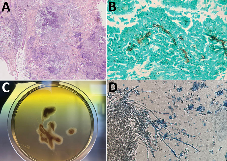
Figure 2. Histological analyses of cutaneous pythiosis in 2 dogs, Italy. A) Section from punch biopsy of cutaneous mass showing confluent pyogranulomatous and eosinophilic granulomas with central necrosis in the deep dermis and panniculus. Hematoxylin and eosin stain; original magnification ×100. B) Section from punch biopsy of cutaneous mass showing broad, irregular branching hyphae. Grocott methenamine silver stain; original magnification ×400. C) Culture (24-hour) of an aspirate from a mass found in case 2. Aspirate was cultured on Sabouraud dextrose agar without antimicrobial drugs, and showed submerged, colorless colonies with irregular radiate patterns. D) Microscopic appearance of the colonies indicating broad (4–10 µm in diameter), hyaline, and sparsely septate hyphae. Original magnification ×250.