Volume 29, Number 9—September 2023
Dispatch
Human Neural Larva Migrans Caused by Ophidascaris robertsi Ascarid
Figure 1
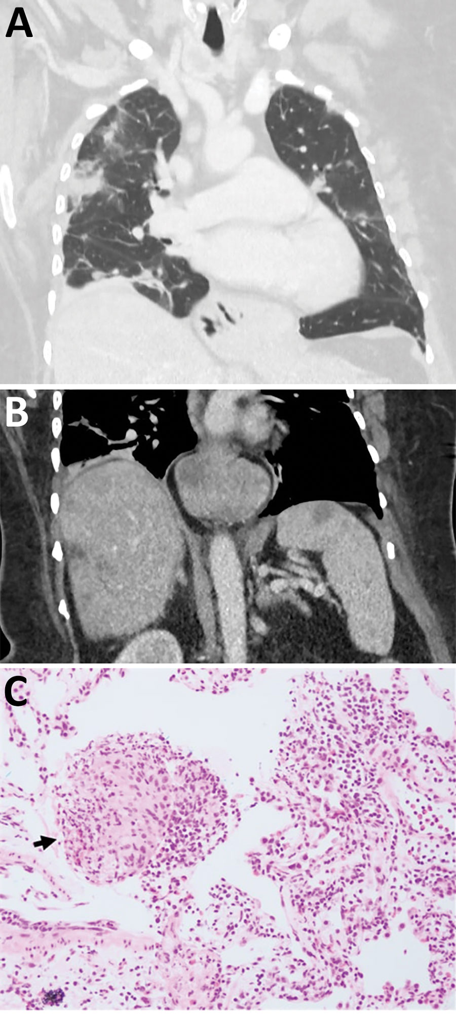
Figure 1. Early testing conducted during investigation of illness in a 64-year-old woman from southeastern New South Wales, Australia, who was later determined to have Ophidascaris robertsi nematode infection. A) Computed tomography scan of chest with venous contrast demonstrating multiple bilateral airspace opacities and nodules with a peripheral bronchovascular distribution. The opacities have surrounding ground-glass changes. Many were present in the patient’s study from a previous hospitalization; however, some had resolved while others were new, indicating a migratory pattern. B) Computed tomography scan of abdomen with venous contrast demonstrating multiple ill-defined hypoattenuated lesions within the liver and spleen. C) Hematoxylin and eosin stain (original magnification ×200) of a pulmonary lesion revealing prominent eosinophil infiltration of stroma and vessel walls. Arrow indicates a granuloma composed of histiocytes and eosinophils. The prominent eosinophilia was inconsistent with hypersensitivity pneumonitis, and the absence of vessel wall damage did not support a diagnosis of eosinophilic granulomatosis with polyangiitis.