Volume 29, Number 9—September 2023
Dispatch
Human Neural Larva Migrans Caused by Ophidascaris robertsi Ascarid
Mehrab E Hossain, Karina J. Kennedy, Heather L. Wilson, David Spratt, Anson Koehler, Robin B. Gasser, Jan Šlapeta, Carolyn A. Hawkins, Hari Priya Bandi, and Sanjaya N. Senanayake
Figure 2
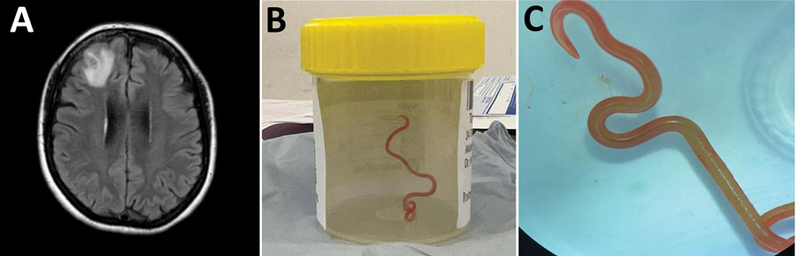
Figure 2. Detection of Ophidascaris robertsi nematode infection in a 64-year-old woman from southeastern New South Wales, Australia. A) Magnetic resonance image of patient’s brain by fluid-attenuated inversion recovery demonstrating an enhancing right frontal lobe lesion, 13 × 10 mm. B) Live third-stage larval form of Ophidascaris robertsi (80 mm long, 1 mm diameter) removed from the patient’s right frontal lobe. C) Live third-stage larval form of O. robertsi (80 mm long, 1 mm diameter) under stereomicroscope (original magnification ×10).
Page created: July 27, 2023
Page updated: August 20, 2023
Page reviewed: August 20, 2023
The conclusions, findings, and opinions expressed by authors contributing to this journal do not necessarily reflect the official position of the U.S. Department of Health and Human Services, the Public Health Service, the Centers for Disease Control and Prevention, or the authors' affiliated institutions. Use of trade names is for identification only and does not imply endorsement by any of the groups named above.