Volume 3, Number 2—June 1997
Synopsis
Japanese Spotted Fever: Report of 31 Cases and Review of the Literature
Cite This Article
Citation for Media
Abstract
Spotted fever group (SFG) rickettsioses, which are transmitted by ticks, were long thought not to exist in Japan. Three clinical cases of Japanese spotted fever (JSF) were first reported in 1984. The causative agent was isolated and named Rickettsia japonica. Through October 1996, 31 cases were diagnosed as JSF in Tokushima Prefecture. Infected patients typically had acute high fever, headache, and characteristic exanthema; eschar was observed in 90%. After the discovery of JSF, more than a hundred cases were reported in southwestern and central Japan. Recent surveys show ticks to be the most probable vectors. As an emerging infectious disease, JSF is not commonly recognized by clinicians; therefore, even though it has not caused fatal cases, it merits careful monitoring.
The spotted fever group (SFG) rickettsioses, which are transmitted by ticks, have a worldwide distribution. Japanese spotted fever (JSF) is one of the newcomers of this group (1); the first clinical cases were reported in 1984 (2). The causative agent was isolated and named Rickettsia japonica (3). Because outbreaks were sporadic and limited, clinical reports concerning JSF, especially from specialists in dermatology and physiology and from general practitioners, were scarce. JSF was first found in Tokushima Prefecture, on the island of Shikoku in southwestern Japan; Tsutsugamushi disease, an important rickettsiosis in Japan, was found there soon afterwards (4). Through October 1996, 31 clinical cases of JSF and 11 cases of Tsutsugamushi disease were diagnosed at Mahara Hospital, Tokushima Prefecture. During the same period, 45 cases of human tick bites were recorded in this JSF-endemic area in the same hospital. This article describes JSF's history, clinical characteristics, and differences from Tsutsugamushi disease and summarizes current information about the epidemiology, vectors, and causative agent of JSF.
In the 1980s, clinicians believed that Tsutsugamushi disease (scrub typhus) was the only rickettsial disease in Japan except for sporadic outbreaks of epidemic typhus in the 1950s. In Tokushima Prefecture, neither disease has been reported in the last two decades. In May 1984, a 63-year-old woman (the wife of a farmer) was hospitalized at Mahara Hospital with high fever and erythematous nonpruritic skin eruptions over the entire body. Antibiotics (ß-lactam and aminoglycoside) used for common febrile infections were not effective, but the patient gradually became afebrile in 2 weeks without effective treatment. In May and July 1984, two additional patients with similar symptoms were treated at the same hospital. Doxycycline was markedly effective in these cases. Before the onset of illness, the patients had collected shoots from bamboo plantations on the same mountain. In two of the patients, an eschar was observed. Tsutsugamushi disease was suspected. However, results of Weil-Felix tests showed positive OX2 serum agglutinins, and OXK were negative in all three cases. These results did not indicate Tsutsugamushi disease, but rather OX2-positive infections, i.e., SFG rickettsioses (1).
The cases were subsequently confirmed by complement fixation test with antigens of SFG rickettsiae (5,6). The name Japanese spotted fever was proposed for these infections (7) and has been commonly used since then (8-10). Oriental spotted fever (11) is a synonym for JSF. The causative agent was isolated in 1986 (12) and named R. japonica (3).
Figure 1

Figure 1. Fever and clinical course, 62-year-old woman.
Figure 2
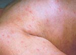
Figure 2. Skin eruptions, hospital day 3.
Figure 3
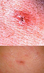
Figure 3. Upper: Typical eschar of Japanese Spotted Fever. Lower: Small and shallow eschar on admission, which disappeared in a few days.
The clinical features of the 31 patients whose illness was diagnosed as JSF at Mahara Hospital from 1984 to October 1996 were analyzed. The disease developed abruptly, with the common symptoms of headache (25 [80%] of 31 patients), fever (31 [100%] of patients), and shaking chills (27 [87%]). Other major objective symptoms of JSF included skin eruptions (31 [100%]) and tick bite eschars (28 [90%]). Most patients (28 [90%]) complained of malaise; joint and muscle pain or numbness of the extremities was rarely mentioned. In the acute stage, remittent fever accompanied by shaking chills was frequently observed. In severe cases, high fever (40°C or more) continued for several days (Figure 1). The maximum body temperature was 38.5°C to 40.8°C (mean 39.5°C), which was higher than that seen in patients with Tsutsugamushi disease (38.5°C ~ 39.1°C). With abrupt high fever or, a few days after onset, fever of unknown origin, the characteristic erythemas developed on the extremities and spread rapidly (in a few hours) to all parts of the body including palms and soles, without accompanying pain or itching. These eruptions were the size of a grain of rice or soybean, and the margin of each of the spots was unclear (Figure 2). The erythemas became remarkable during the febrile period and tended to spread more over the extremities than the trunk. Palmar erythema, a characteristic finding not seen in Tsutsugamushi disease, disappeared in the early stage of the disease. The erythemas became petechial after 3 to 4 days, peaked in a week or 10 days, and disappeared in 2 weeks. However, in severe petechial cases, the brown pigmentation remained for 2 months or more. Eschar was observed on the hands, feet, neck, trunk, and shoulders of patients (Figure 3). This eschar generally remained for 1 to 2 weeks, but in some cases it disappeared in a few days. Eschars in JSF patients are smaller than those seen in patients with Tsutsugamushi disease and may be missed without careful observation. Regional or generalized lymphadenopathy, which is observed in almost all cases of Tsutsugamushi disease, was not remarkable in JSF patients. Swelling of the liver and spleen was observed in a few patients. One patient had cardiomegaly (5), and in another area, a patient had central nervous system involvement (13).
The results of laboratory examinations of JSF patients are almost the same as those of patients with common SFG rickettsioses. During clinical examinations at the initial stage of the disease, urinalyses registered a slight positive reading for protein and occult blood, which may lead to a misdiagnosis of urinary infection. In the acute stage, leukocytosis may also be found with leukopenia (3,600~12,800), and a left shift in leukocyte count was observed. Thrombocytopenia (6.8~35.3) may also be found. In week 1 to 2, leukocyte counts increased slightly, and lymphocyte counts tended to increase. Among biochemical examinations, C-reactive proteins were strongly positive, and liver functions were slightly impaired but returned to normal in 2 to 3 weeks.
Serologic Results
Serodiagnosis for JSF is usually performed by the indirect immunoperoxidase (IP) or immunofluorescence (IF) techniques, with antigens prepared from R. japonica or other SFG rickettsiae. With the IP test, IgG and IgM antibodies were detected in the sera beginning on day 9 after the onset of fever; titers of IgG antibodies were higher than those of IgM antibodies (14). The IF test had similar results (15). In the 31 clinically diagnosed cases of JSF in Tokushima Prefecture at Mahara Hospital, all patients had significant changes in serum IP antibody titers to R. japonica, and 27 (87%) had significant changes in OX2 agglutinin titers by the Weil-Felix technique (10,14; F. Mahara, unpub. data).
Treatment
Antibiotics such as penicillins, ß-lactams, or aminoglycosides, commonly used in the empiric treatment of febrile disease, were completely ineffective, but doxycycline and minocycline were markedly effective in treating the JSF patients (16).
On the first day of hospitalization in one patient with severe disease (Figure 1), fever of more than 40°C with shaking chills persisted after the antibiotics cefazolin and fosfomycin were administered. The general condition of the patient worsened and on the morning of the third day, generalized edema and confusion developed. On day 3, the patient was given drip infusion of doxycycline 300 mg per day, which was dramatically effective; the fever decreased to 38°C during the first drip infusion.
In an in vitro study, minocycline was the most effective antibiotic against R. japonica, followed by other tetracycline antibiotics (17,18). In contrast, the sensitivity to ß-lactam and penicillin was lower or negligible, but quinolones were effective against the JSF agent (18). Three patients were treated with a new quinolone (tosufloxacin 300 mg per day in three divided doses per os), which proved effective in two cases (9). Patients with dehydration received drip infusion of doxycycline or minocycline (200 mg to 300 mg per day for 3 to 7 days), and after becoming afebrile, received 200 mg per day 2 divided doses to prevent relapse. Patients suspected of having JSF should receive empiric therapy with minocycline or doxycycline without waiting for serologic confirmation of illness.
Figure 4

Figure 4. The number of Japanese spotted fever patients in Japan (1984-1995).
Figure 6

Figure 6. Seasonal prevalence of Japanese spotted fever and Tsutsugamushi disease.
Figure 5

Figure 5. Geographic distributions of JSF and Tsutsugamushi disease in Japan.
From 1984 to 1995, 144 cases of JSF were reported by the National Institutes of Health in Japan (19; Figure 4). Case reports included only the number of cases and prefectures where cases were reported, including Tokushima Prefecture. According to this information, JSF-endemic prefectures are located along the coast of southwestern and central Japan in a warm climate (Figure 5). The landscape is diverse, including bamboo plantations, crop fields, coastal hills, and forests (20). Of the 31 JSF patients in Tokushima Prefecture, nine were male, and 22 were female. Ages were 4 to 78 years, but most patients (68%) were 60 to 70 years old. The onset of the disease was 2 to 8 days after work in the fields. In this prefecture, farmers work in the forest to gather bamboo shoots in the spring and chestnuts in the autumn; cases occurred from April to October (6,18; Figure 6). In this area, we can distinguish seasonal differences in the occurrence of JSF and Tsutsugamushi disease; whereas JSF occurs from spring to autumn, Tsutsugamushi disease occurs in winter (November-February). Although seasonal differences occur in the western parts of Japan, the prevalent seasons for Tsutsugamushi disease vary in other parts of Japan (21).
Like other SFG rickettsioses, JSF is presumed to be transmitted by a tick bite. The high proportion of patients with tick bite eschars supports this hypothesis. Thirteen JSF patients (28.9%) recalled tick bites before the onset of illness; however, the ticks had been lost, and no specimens from the patients were available for further study. From 1984 to October 1996, 45 persons with tick bites were recorded at Mahara Hospital (Table 1). Identified ticks included three genera and eight species.
Three genera and six species of ticks have been reported as positive for R. japonica in JSF-endemic areas (Table 2). Hemolymph samples from Dermacentor taiwanensis, Haemaphysalis flava, Haemaphysalis formosensis, Haemaphysalis hystricis, Haemaphysalis longicornis, and Ixodes ovatus were positive when tested by the IP technique using a species-specific monoclonal antibody against R. japonica (22). R. japonica was also detected by IF in the hemolymph of H. longicornis (23). A polymerase chain reaction (PCR) technique using species-specific primers detected R. japonica in H. hystricis (24), H. flava, and I. ovatus (25). The agent has also been detected in H. longicornis by restriction fragment length polymorphism of PCR product (23). Of these, H. flava, H. longicornis, and I. ovatus commonly feed on humans in Japan (26). Recent tick surveys in Japan have identified two serotypes or species of SFG rickettsial isolates other than R. japonica, which are of uncertain clinical significance (27,28).
The etiologic agent was first isolated from a patient (in Kochi Prefecture) in 1986 (12). In 1987, the causative rickettsia was also isolated from a JSF patient in Tokushima Prefecture (29-31). The former isolate is the type strain YH (ATCC VR-1363), later named R. japonica, a new SFG rickettsia; the latter strain (Katayama) was the first isolate from JSF-endemic areas outside Kochi (3,32). The Katayama strain type and R. japonica were demonstrated by serologic analysis using monoclonal antibodies (33). Serotyping by use of the reciprocal crossreactions of mouse antisera to six human isolates from Tokushima and the type strain YH or R. japonica also indicated that these are the same species (34). In 1988, another strain was isolated from a patient in Awaji Island, Hyogo Prefecture, which is considered a new area of JSF-endemic disease (35).
Recently, an isolate from a febrile patient in Wakayama Prefecture was also reported as R. japonica (36). In an electron microscopy study, R. japonica were generally recognized as short rods or pleomorphic coccobacillary forms less than 2 mm in length and 0.5 mm in diameter and could be found not only in the cytoplasms but also in the nuclei of the host cells (37). A multilayered mesosome-like structure was observed in the rickettsiae multiplying in a host cell (38). This unique structure has not been reported in other species except in Rickettsia prowazekii (39). After the initial isolation, at least 20 rickettsial strains have been isolated from JSF patients by cell culture techniques or nude mouse passage in Tokushima, Kochi, Hyogo, Chiba, and Wakayama Prefectures. However, it has not been determined if their strains differ in virulence.
Ten diseases caused by SFG rickettsiae have been reported in humans (40): Rocky Mountain spotted fever, Mediterranean spotted fever, Siberian tick typhus, African tick bite fever, Queensland tick typhus, Japanese spotted fever, Israeli spotted fever, Astrakhan spotted fever, Flinders Island spotted fever, and rickettsialpox. The clinical symptoms of JSF—a triad of high fever, skin eruptions, and tick bite eschar—are similar to those of typical SFG rickettsioses. With regard to skin eruptions, eschar, and severity of the disease, JSF is more akin to Mediterranean spotted fever and Siberian tick typhus than to Rocky Mountain spotted fever. Recent tick surveys indicated that the most probable vectors of JSF are H. flava, H. longicornis, and I. ovatus.
In Japan, the clinical features of JSF are similar to those of Tsutsugamushi disease; however, close clinical observation exposes the differences between the two diseases. Widespread outbreaks of Tsutsugamushi disease have been reported repeatedly in recent years (21). No fatal cases of JSF have been reported. However, death rates from other SFG rickettsioses (approximately 2.5% for Mediterranean spotted fever [41] and 3% to 7% from Rocky Mountain spotted fever [42]) suggest that unless JSF is treated appropriately, it can pose the same risk. If JSF is suspected, empiric treatment should begin without delay during the early stages of disease.Note: This article is also available in Japanese.To access the Japanese Translation of this article, a browser that supports Japanese is required.
Dr. Mahara is chief director of Mahara Hospital, Tokushima, Japan. He serves as a counselor of the Japanese Association for Infectious Diseases and is a member of the International Eurasian Academy of Sciences.
Acknowledgment
The author thanks Professor Emeritus Suto T., Department of Microbiology, Akita University, for serologic confirmation; Professor Jan Kazar, Slovak Academy of Sciences, for his helpful advice; Drs. Fujita H., Ohara Research Laboratory and Takada N., Fukui Medical School, for epidemiologic studies; and all colleagues who collaborated in the investigations.
References
- Brouqui P, Raoult D. Clinical aspect of human SFG rickettsiae infection in the era of molecular biology. In: Kazar J, Toman R, editors. Proceedings of the 5th International Symposium on Rickettsiae and Rickettsial Diseases. Bratislava, Slovak: Slovak Academy of Science, 1996;195-210.
- Mahara F. Three Weil-Felix reaction (OX2) positive cases with skin eruptions and high fever. Journal of Anan Medical Association. 1984;68:4–7.
- Uchida T, Uchiyama T, Kumano K, Walker DH. Rickettsia japonica sp. nov., the etiological agent of spotted fever group rickettsiosis in Japan. Int J Syst Bacteriol. 1992;42:303–5. DOIPubMedGoogle Scholar
- Mahara F, Fujita H, Suto T. 11 cases of Japanese Spotted Fever and first report of the Tsutsugamushi disease in Tokushima Prefecture. J Jpn Assoc Infect Dis. 1989;63:963–4.
- Mahara F, Koga K, Sawada S, Taniguchi T, Shigemi F, Suto T, J Jpn Assoc. The first report of the rickettsial infections of spotted fever group in Japan; three clinical cases. Infect Dis. 1985;59:1165–72.
- Mahara F. Clinical pictures of the spotted fever group rickettsiosis. J Jpn Assoc Clin Virol. 1985;13:447–52.
- Mahara F. Japanese spotted fever: a new disease named for spotted fever group rickettsiosis in Japan. Annu Rep Ohara Hosp. 1987;30:83–91.
- Emilio Weiss. Rickettsias. Joshua Lederberg, editor. Encyclopedia of Microbiology, Academic Press, Harcourt Brace Jovanovich, New York, 1992; Vol.3:585-610.
- Mahara F. Japanese Spotted Fever. Oka H, Wada T, editors. Encyclopedia of Medical Science, Kodansya, Tokyo, 1992; suppl.9:27-30.
- Mahara F. Japanese Spotted Fever, clinical features and vectors. Kazar J, Toman R, editors. Proceedings of the Vth International Symposium on Rickettsiae and Rickettsial Diseases. Publishing House of the Slovak Academy of Science, Bratislava, Slovak, 1996;641-6.
- Uchida T. Rickettsia japonica, the etiologic agent of oriental spotted fever. Microbiol Immunol. 1993;37:91–102. PubMedGoogle Scholar
- Uchida T, Tashiro F, Funato T, Kitamura Y. Isolation of a spotted fever group rickettsia from a patient with febrile exanthematous illness in Shikoku, Japan. Microbiol Immunol. 1986;30:1323–6. PubMedGoogle Scholar
- Iwamoto K, Nishimura F, Yoshino Y, Mihara J, Okabe T, Kameda H, J Jpn Assoc. A case of spotted fever with central nervous system involvement. Infect Dis. 1988;62:1192–6
- Amano K, Hatakeyama H, Okuta M, Suto T, Mahara F. Serological studies of antigenic similarity between Japanese spotted fever rickettsiae and Weil-Felix test antigens. J Clin Microbiol. 1992;30:2441–6. PubMedGoogle Scholar
- Uchida T, Tashiro F, Funato T, Kitamura Y. Immunofluorescence test with Rickettsia montana for serologic diagnosis of rickettsial infection of the spotted fever group in Shikoku, Japan. Microbiol Immunol. 1986;30:1061–6. PubMedGoogle Scholar
- Mahara F. Clinical findings of Japanese Spotted Fever and Tsutsugamushi disease. Acari-Disease Interface, (ed.) Organizing Committee of SADI, Yuki Press Inc., Fukui, 1994;38-45.
- Suto T, Hatakeyama H, Ito R, Nakamura Y. J Jpn Assoc. In vitro susceptibility of a strain of rickettsia recently isolated from a case of Japanese spotted fever to chemotherapeutic agents. Infect Dis. 1989;63:35–8.
- Miyamura S, Oota T. In vitro susceptibility of rickettsial strains from patients with Japanese Spotted Fever to quinolones, penicillins and other selected chemotherapeutic agents. Chemotherapy. 1991;39:258–60.
- Tsuboi Y. The laboratory examination method for rickettsioses. Clin Virol. 1995;23:394–9.
- Takada N. Review: recent findings on vector acari for rickettsia and spirochete in Japan. Jap J Sanit Zool. 1995;46:91–108.
- Suto T. Tsutsugamushi disease now. Med J Akita City Hosp. 1995;4:1–18.
- Takada N, Fujita H, Yano Y, Oikawa Y, Mahara F. J Jpn Assoc. Vectors of Japanese spotted fever. Infect Dis. 1992;66:1218–25.
- Uchida T, Yan Y, Kitaoka S. Detection of Rickettsia japonica in Haemaphysalis longicornis ticks by restriction fragment length polymorphism of PCR product. J Clin Microbiol. 1995;33:824–8. PubMedGoogle Scholar
- Furuya Y, Katayama T, Yoshida Y, Kaiho I, Fujita H. Analysis of Rickettsia japonica DNA and detection of the DNA by PCR. Acari-Disease Interface (ed.) Organizing Committee of SADI, Yuki Press Inc., Fukui, 1994;141.
- Katayama T, Furuya Y, Yoshida Y, Kaiho I. J Jpn Assoc. Spotted fever group rickettsiosis and vectors in Kanagawa Prefecture. Infect Dis. 1996;70:561–8.
- Yamaguti N. Human tick bites in Japan. Acari-Disease Interface (ed.) Organizing Committee of SADI, Fukui: Yuki Press Inc., 1994;16-23.
- Fujita H, Watanabe Y, Takada N, Yano Y, Tsuboi Y, Mahara F. Spotted fever group rickettsiae isolated from ticks in Japan. Acari-Disease Interface (ed.) Organizing Committee of SADI, Fukui: Yuki Press Inc., 1994;142-9.
- Takada N, Fujita H, Yano Y, Tsuboi Y, Mahara F. First isolation of a rickettsia closely related to Japanese spotted fever pathogen from a tick in Japan. J Med Entomol. 1994;31:183–5. PubMedGoogle Scholar
- Fujita H, Watanabe Y, Mahara F. Isolation of a causative rickettsia from a patient with Japanese spotted fever in Tokushima Prefecture, Japan. Annu Rep Ohara Hosp. 1988;31:17–21.
- Kobayashi Y, Tange Y, Kanemitsu N, Okada T, Mahara F. J Jpn Assoc. The causative agent from a patient with spotted fever group rickettsiosis in Tokushima Japan. Infect Dis. 1988;62:1132–7.
- Hatakeyama H, Ito R, Nakamura Y, Suto T, Amano K, Mahara F. Characterization of spotted fever group rickettsiae isolated from Japanese spotted fever patients. Clinical Microbiology. 1991;18:103–8.
- Uchida T, Yu X, Uchiyama T, Walker DH. Identification of a unique spotted fever group rickettsia from humans in Japan. J Infect Dis. 1989;159:1122–6. PubMedGoogle Scholar
- Oikawa Y, Takada N, Fujita H, Yano Y, Tsuboi Y, Ikeda T. Identity of pathogenic strains of spotted fever rickettsiae in Shikoku district based on reactivities to monoclonal antibodies. Jpn J Med Sci Biol. 1993;46:45–9. PubMedGoogle Scholar
- Fujita H, Watanabe Y, Takada N, Tsuboi Y, Mahara F. Isolation and serological identification of causative rickettsiae from Japanese spotted fever patients. Asian Med J. 1993;36:660–5.
- Kobayashi Y, Tange Y, Okada T, Kodama K. J Jpn Assoc. The causative agent from a patient with spotted fever group rickettsiosis in Japan on Awaji Island, Hyogo. Infect Dis. 1990;64:413–8.
- Fujita H. Isolation of Rickettsia japonica from a febrile patient-Wakayama. Infectious Agents Surveillance Report. 1995;16:30.
- Iwamasa K, Okada T, Tange Y, Kobayashi Y. Ultra-structural study of the response of cells infected in vitro with causative agent of spotted fever group rickettsiosis in Japan. APMIS. 1992;100:535–42. DOIPubMedGoogle Scholar
- Amano K, Hatakeyama H, Sasaki Y, Ito R, Tamura A, Suto T. Electron microscopic studies on the in vitro proliferation of spotted fever group rickettsia isolated in Japan. Microbiol Immunol. 1991;35:623–9.PubMedGoogle Scholar
- Silverman DJ, Wisseman CL Jr. Comparative ultra-structural study on the cell envelopes of Rickettsia prowazekii, Rickettsia rickettsii, and Rickettsia tsutsugamushi. Infect Immun. 1978;21:1020–3. PubMedGoogle Scholar
- Beati L, Raoult D. Spotted Fever Rickettsiae. Kazar J, Toman R, editors. Proceedings of the Vth International Symposium on Rickettsiae and Rickettsial Diseases. Publishing House of the Slovak Academy of Science, Bratislava, Slovak 1996;134-69.
- Raoult D, Weiller PJ, Chagnon A, Chaudet H, Gallais H, Casanova P. Mediterranean spotted fever . Am J Trop Med Hyg. 1986;35:845–50.PubMedGoogle Scholar
- Hattwick MAW, O'Brien RJ, Hanson B. Rocky mountain spotted fever: epidemiology of increasing problem. Ann Intern Med. 1986;84:732–9.
Figures
Tables
Cite This ArticleTable of Contents – Volume 3, Number 2—June 1997
| EID Search Options |
|---|
|
|
|
|
|
|