Volume 3, Number 2—June 1997
Synopsis
Polycystic Kidney Disease: An Unrecognized Emerging Infectious Disease?
Figure 2
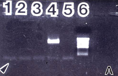
Figure 2. Amplification results of normal and PKD kidney tissue and cyst fluids with universal fungal primers ITS 1 and NL 4. 2A: DNA from healthy human kidney tissue diluted 1:10, 1:100 and 1:1,000 (lanes 1-3); control fungal DNA, A. tamarii (lane 4); negative control (lane 5); 1 kb ladder (lane 6); arrow indicates migration front. 2B (NL 4) and 2C (ITS 1): two cyst fluids, donor 6, negative for detectable endotoxin and ß-DG (lanes 1 and 5); two cyst fluids, donor 4, positive for ß-DG (lanes 2 and 3) and positive for Fusarium solani antigen (lane 3); two cyst fluids, donor 5, positive for ß-DG (lanes 4 and 6); two PKD kidney tissues, donor 5 (lane 7) and donor 4 (lane 8); negative control (lane 9). Large arrows in 2B and 2C point to 560 bp molecular weight marker; small arrows point to product bands.