Volume 30, Number 1—January 2024
Dispatch
Hantavirus Disease Cluster Caused by Seoul Virus, Germany
Cite This Article
Citation for Media
Abstract
A cluster of 3 persons in Germany experienced hantavirus disease with renal insufficiency. Reverse transcription PCR–based genotyping revealed infection by Seoul hantavirus transmitted from pet rats. Seoul virus could be responsible for disease clusters in Europe, and infected pet rats should be considered a health threat.
Hantavirus disease occurs worldwide. Estimated number of cases is ≈100,000 annually; most cases are in Asia and Europe. Pathogenic hantaviruses are transmitted to humans through excreta of infected rodents. The infection can lead to renal or cardiopulmonary failure; case-fatality rates are up to 50% (1). Seoul virus (SEOV) is a hantavirus species for which rats (Rattus spp.) are natural hosts. In humans, SEOV infection causes mild to moderate disease with fever, acute kidney injury, hepatitis, and gastroenteritis; it is associated with transient thrombocytopenia and proteinuria (2–4).
Despite numerous clinical cases and local outbreaks of SEOV infection in Asia, few human cases are known in Europe (4–6). However, SEOV infections are difficult to identify by routine serodiagnosis because of high cross-reactivity with antigens of related hantaviruses, such as Hantaan virus and Dobrava-Belgrade virus. Whereas Hantaan virus is endemic in Asia, Dobrava-Belgrade virus is found in many parts of Europe (1).
Autochthonous human SEOV infection in Germany was described in 2020; the hantavirus type was determined by molecular analysis, and infection source was identified as a pet rat (4). Deeper molecular analysis found SEOV strains in several pet rats, including the animals owned by that patient (7). However, no clusters of human SEOV infections were reported previously from Germany or any other part of Europe.
During November–December 2021, three persons in Germany experienced typical initial signs of hantavirus disease (fever, malaise, myalgia, chills, low-back pain, nausea) (Table 1). The patients lived ≈25 km apart. All 3 patients had acute kidney injury with reduction of glomerular filtration rate, proteinuria, and microhematuria. Moreover, biochemical analysis revealed typical thrombocytopenia and signs of inflammation, including elevation of C-reactive protein and transaminases. Serum creatinine levels remained within reference ranges or were only transiently higher. The patients recovered and were discharged after 3–6 days (Table 1).
All 3 patients developed IgM and IgG against diagnostic antigens from the group of murine-associated hantaviruses, including Hantaan virus, Dobrava-Belgrade virus, and SEOV. After onset of disease, we performed panhantavirus PCR, reverse transcription PCR based on amplification of a 412-nt region of the genomic large segment (8), to detect hantavirus genetic material from serum samples of patients 1 and 3 (Table 2).
Patients 1 and 2 were married to each other and lived in the federal state of North Rhine-Westphalia. Their children (persons 12a and 12b) (Table 2) remained healthy and did not seroconvert. The family was known to keep pet rats at home. Patient 3, who lived in Lower Saxony, reported that his girlfriend also kept pet rats. Investigation of his girlfriend (person 4) showed that she remained healthy; however, her hantavirus IgM negative/IgG positive serostatus suggested a previous subclinical infection (Table 2).
Figure
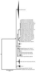
Figure. Phylogenetic tree of partial large segment Seoul virus sequences from humans and rats, Germany. Segments were 412-nt long, positions nt 2919–3330 based on reference sequence (KM948594_Cherwell_GBR_BR). The partial large segment...
The family of patients 1 and 2 permitted us to test their pet rats. We amplified the corresponding region from the genomic large segment. Phylogenetic analysis of the amplified virus sequences from patients 1 and 3 and the pet rats revealed almost identical sequences (Figure). We compared them to the amplified virus sequences from a previous unrelated patient and her pet rat from Lower Saxony (4).
Phylogenetic analysis disclosed the detected virus sequences as SEOV and, moreover, demonstrated the high relatedness of virus sequences from patient and pet rats from North-West Germany. The high similarity of SEOV sequences from Germany to sequences derived from breeder rats in the Netherlands, France, and the United States, but not to sequences from wild rats of those countries, suggests an intensive exchange of pet rats between neighboring countries in Europe and between Europe and the United States (7).
Human SEOV infections might be underestimated because the nucleocapsid proteins used in serologic assays share high amino acid sequence similarity and cross-reactivity with different hantavirus species. To explore this hypothesis in an external quality assessment, we sent a serum sample from patient 3, obtained 2 months after complete recovery, to 8 expert laboratories in Europe. All laboratories analyzed the sample in their routine hantavirus diagnostics, including commercial and lab-derived enzyme immunoassays, immunofluorescence assays, rapid assays, and immunoblots for confirmation (Appendix Tables 1, 2). Results revealed strong cross-reactivities to related hantavirus nucleocapsid proteins in IgG and IgM screening and in confirmation assays, making correct serotyping impossible. Thus, in cases of unexpected pattern in serodiagnostics or cases of known contact to rats, we strongly recommend verifying positive results with molecular methods or by typing of neutralizing antibodies. Only the laboratory using a focus reduction neutralization assay identified the SEOV infection (Appendix Table 1).
A total of 6 molecularly proven SEOV-infected patients in Germany have been described: 1 imported case (9), 1 autochthonous case (4), and the 3 symptomatic case-patients and 1 asymptomatic case-patient we described in this report. Five of these persons had been admitted to hospital for several days. In all autochthonous cases, pet rats were confirmed as source of infection, strongly suggesting a need for close cooperation between public health and animal health institutions in the One Health frame. Pet rats, in addition to wild and breeder or feeder rats, should be considered threats for SEOV infection in humans.
Dr. Hofmann is the chair of the National Consultation Laboratory for Hantaviruses, Institute of Virology, Charité University Medicine, Berlin, Germany. His primary research interest is human infections with viral pathogens.
Acknowledgments
We thank J. Dreesman, C. Klier, and M. Oskamp from local health authorities for their support. We thank the 8 expert laboratories in Europe for analyzing the serum sample, as described in the Appendix Tables. We thank M. Raftery for critical reading of the manuscript and C. Stephan for expert technical assistance.
This work was supported by the German Centre for Infection Research, thematic translational unit Emerging Infections (grant no. 01.808 00) awarded to R.G.U.
References
- Kruger DH, Figueiredo LT, Song JW, Klempa B. Hantaviruses—globally emerging pathogens. J Clin Virol. 2015;64:128–36. DOIPubMedGoogle Scholar
- Lee HW. Hemorrhagic fever with renal syndrome in Korea. Rev Infect Dis. 1989;11(Suppl 4):S864–76. DOIPubMedGoogle Scholar
- Clement J, LeDuc JW, McElhinney LM, Reynes JM, Van Ranst M, Calisher CH. Clinical characteristics of ratborne Seoul hantavirus disease. Emerg Infect Dis. 2019;25:387–8. DOIPubMedGoogle Scholar
- Hofmann J, Heuser E, Weiss S, Tenner B, Schoppmeyer K, Esser J, et al. Autochthonous ratborne Seoul virus infection in woman with acute kidney injury. Emerg Infect Dis. 2020;26:3096–9. DOIPubMedGoogle Scholar
- Zhang YZ, Dong X, Li X, Ma C, Xiong HP, Yan GJ, et al. Seoul virus and hantavirus disease, Shenyang, People’s Republic of China. Emerg Infect Dis. 2009;15:200–6. DOIPubMedGoogle Scholar
- Clement J, LeDuc JW, Lloyd G, Reynes JM, McElhinney L, Van Ranst M, et al. Wild rats, laboratory rats, pet rats: global Seoul hantavirus disease revisited. Viruses. 2019;11:652. DOIPubMedGoogle Scholar
- Heuser E, Drewes S, Trimpert J, Kunec D, Mehl C, de Cock MP, et al. Pet rats as the likely reservoir for human Seoul orthohantavirus infection. Viruses. 2023;15:467. DOIPubMedGoogle Scholar
- Klempa B, Fichet-Calvet E, Lecompte E, Auste B, Aniskin V, Meisel H, et al. Hantavirus in African wood mouse, Guinea. Emerg Infect Dis. 2006;12:838–40. DOIPubMedGoogle Scholar
- Hofmann J, Weiss S, Kuhns M, Zinke A, Heinsberger H, Kruger DH. Importation of human Seoul virus infection to Germany from Indonesia. Emerg Infect Dis. 2018;24:1099–102. DOIPubMedGoogle Scholar
Figure
Tables
Cite This ArticleOriginal Publication Date: December 15, 2023
Table of Contents – Volume 30, Number 1—January 2024
| EID Search Options |
|---|
|
|
|
|
|
|
Please use the form below to submit correspondence to the authors or contact them at the following address:
Jörg Hofmann, Charité Medical School—Institute of Medical Virology, Chariteplatz 1 Berlin 10117, Germany
Top