Volume 30, Number 11—November 2024
Dispatch
Genomic Epidemiology of Human Respiratory Syncytial Virus, Minnesota, USA, July 2023–February 2024
Cite This Article
Citation for Media
Abstract
We recently expanded the viral genomic surveillance program in Minnesota, USA, to include human respiratory syncytial virus. We performed whole-genome sequencing of 575 specimens collected at Minnesota healthcare facilities during July 2023–February 2024. Subgroups A and B differed in their genomic landscapes, and we identified 23 clusters of genetically identical genomes.
Human respiratory syncytial virus (RSV) is a major respiratory pathogen with increased risk of severe infections among infants and young children, elderly persons, and persons with underlying health conditions, including immunocompromise (1). Investigators have applied whole-genome sequencing (WGS) for retrospective RSV surveillance and outbreak investigation in the United States (2), but WGS has not yet been documented as a tool for prospective surveillance. Although researchers have developed and improved several genetic typing schemes to facilitate more granular characterization of RSV (3), application of those schemes in genomic epidemiology has not been thoroughly evaluated. We report preliminary findings from our recently established genomic surveillance program for RSV in Minnesota, USA. Because this study was conducted as a component of public health surveillance subject to Minnesota Reporting Rules, our investigation required no institutional review board approval.
During July 2023–February 2024, we collected RSV-positive nasal or nasopharyngeal specimens submitted voluntarily from outpatients and inpatients tested by quantitative reverse transcription PCR (qRT-PCR) or rapid antigen detection assays in 11 healthcare facilities in Minnesota. Specimens arrived with limited patient data (name, sex assigned at birth, date of birth, date of specimen collection, and outpatient or inpatient status).
We amplified genomes from specimens using 50 pairs of PCR primers that generated overlapping amplicons, as described by Maloney et al. (unpub. data, https://virological.org/t/preliminary-results-from-two-novel-artic-style-amplicon-based-sequencing-approaches-for-rsv-a-and-rsv-b/918), and sequenced genomes using the GridION platform with R9 flow cell chemistry (Oxford Nanopore Technologies, https://nanoporetech.com). We performed genome assembly, quality control, and viral subtyping using the nf-core Viralrecon pipeline (4), using a quality threshold of 20-fold coverage over 90% of the viral genome. We used Nextclade software to subgroup genomes based on the whole-genome lineage typing scheme described by Goya et al. (3) as available through Nextclade in June 2024 (5).
We successfully sequenced 575 RSV genomes from this cohort of case-patients. Median patient age was 2 years; 91.8% were <18 years of age, and 5% were ≥65 years of age. Patient sex was 53.4% male and 46.6% female. We classified 287 (49.9%) genomes as subgroup A and 288 (50.1%) as subgroup B (Table 1). Most RSV-A genomes (98.9%; n = 284) were distributed among 4 whole-genome lineages: A.D.1 (10.8%; n = 31), A.D.3 (18.5%; n = 53), A.D.5 (38.0%; n = 109), and A.D.5.2 (31.7%; n = 91). By contrast, most RSV-B genomes (95.1%; n = 274) belonged to a single lineage, B.D.E.1.
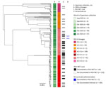
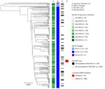
We constructed phylogenetic trees using Nextstrain’s Augur pipeline version 24.3.0 (Figures 1, 2) (6,7). We aligned viral genome assemblies to Nextstrain’s default reference sequences (hRSV/A/England/397/2017 and hRSV/B/Australia/VIC-RCH056/2019) by using MAFFT version 7.526 (8) and then constructed and refined distance-scaled and time-scaled trees from those alignments by using IQTree version 2.3.3 and TimeTree version 0.11.3 (9,10). Comparisons of tree architecture showed greater diversity among all RSV-A genomes (mean pairwise p-distance 0.00933) than among all RSV-B genomes (mean pairwise p-distance 0.00455) (11). We observed pairwise p-distances among lineages with >3 genomes to be more comparable across subgroups (Table 1). Time-scaled phylogenetic analyses specific to the genomes we sequenced estimated the divergence of lineages to have occurred between 2 and 8 years before their earliest specimen collection dates (Table 1; Appendix Figure). The 95% CIs excluded divergence dates that were more recent than 1–5 years before earliest specimen collection.
We identified single-nucleotide polymorphisms (SNPs) from reference-based whole-genome alignments using SNP-dists version 0.8.2 (12). Pairwise comparisons showed that 32.3% of genomes were identical to >1 other genome at 0 SNPs (RSV-A, 33.7%, n = 97; HRSV-B, 30.9%, n = 89) and 53% within 1 SNP (RSV-A, 54%, n = 155; RSV-B, 52.1%, n = 150). We found 23 clusters of at least 3 genomes with shared nucleotide identity at 0 SNPs (RSV-A, n = 14; RSV-B, n = 9). Those clusters included 19.5% of all genomes (RSV-A, 20.6%, n = 59; RSV-B, 18.4%, n = 53). Additionally, we identified 1 clade of 8 RSV-B genomes with the F protein amino acid mutation K68N, which is associated with nirsevimab resistance (13). This clade had an estimated time of most recent common ancestor of September–November 2023 (Figure 2).
To assess our ability to link RSV genomes to known severe infections, we cross-referenced the names and birthdates of case patients whose specimens we sequenced against the Respiratory Syncytial Virus Hospitalization Surveillance Network (RSV-NET). RSV-NET is a component of the Centers for Disease Control and Prevention Emerging Infections Program focused on sentinel population-based surveillance of RSV-associated hospitalizations and deaths (Table 2; Figure 1) (14). Among 531 genomes collected during October 2023–January 2024, the peak months of specimen collection, 117 (22%) were noted among RSV-NET cases (RSV-A, 55.2%, n = 64; RSV-B, 45.3%, n = 53). Those 117 genomes represented 6.3% of all hospitalized cases of RSV during that period. Nine (39.1%) of the 23 clusters we identified included >1 RSV-NET case (RSV-A, 50%, n = 7; RSV-B, 22.2%, n = 2), and 13.4% of clustered cases were in RSV-NET (RSV-A, 20.3%, n = 2; RSV-B, 5.7%, n = 3). Two RSV-NET case-patients with sequenced RSV—both with A.D.5.2 infections—were documented to have received nirsevimab (Figure 1). Of the 8 RSV-B cases whose genomes carried the K68N F protein mutation, 1 was documented in RSV-NET (Figure 2).
To assess potential biases in our sampling for WGS, we performed a preliminary representativeness analysis of hospitalized RSV case-patients with or without collected WGS data. The cohort of sequenced cases documented in RSV-NET was not fully representative of all RSV-NET cases (Table 2). Compared with all RSV-NET case-patients, RSV-NET case-patients with sequenced RSV tended to be younger (median age 2 years for all RSV-NET vs. 1 year for sequenced RSV-NET; p = 0.0193), included a smaller proportion of cases of White versus non-White race (63.6% of all RSV-NET vs. 53.5% for sequenced RSV-NET; p = 0.0014), and were more likely to live within the Minneapolis-St. Paul metropolitan area (65% of all RSV-NET vs. 87.1% of sequenced RSV-NET; p<0.001).
Our findings from a new statewide genomic surveillance program of RSV infections in Minnesota, USA, show that applying WGS for RSV surveillance can yield insights into viral circulation and population dynamics. Prospective sequencing revealed differing genomic landscapes of subgroups A and B and contextualized their genetic diversity within the state. The detection of clusters within the viral population shows the potential use of WGS for detection and investigation of RSV outbreaks at state or local levels. Our study also provided novel cross-referencing of viral genomic data against sentinel clinical surveillance of severe RSV infections.
Our study’s skewed and localized convenience sampling limited the analytical potential of our findings. The limited geographic and temporal scope of our study also potentially introduced variability in our time-scaled analyses and location-specific discrepancies between RSV evolution in Minnesota and in other regions. In future work, we will expand the collection of genomic and epidemiologic data with more targeted collection of specimens, to improve the scope and representativeness of our sampling approach such that these questions can be investigated more thoroughly. We also intend to perform sufficiently powered epidemiologic analyses on the emergence of mutations linked to vaccine escape, virulence, and transmissibility. However, our results demonstrate that prospective genomic surveillance of RSV can identify the emergence of mutations and clades of epidemiologic significance, such as those linked to evolution of vaccines or monoclonal antibody resistance (2,3).
Mr. Evans is a genomic epidemiologist with the Minnesota Department of Health. His work focuses on developing, implementing, and optimizing the use of genomics for pathogen surveillance and epidemiological intervention.
Acknowledgments
We thank the following healthcare facilities for submitting specimens: Carris Rice, CCM Health-Montevideo, Children’s Minnesota, Fairview Ridges, Fairview Southdale, Grand Itasca, Hennepin County Medical Center, Maple Grove Hospital, the Hennepin County medical examiner’s office, Minnesota State University-Mankato clinic, Ridgeview-Waconia, and Riverview-Crookston. We thank the Centers for Disease Control and Prevention RSV-NET program and the Emerging Infections Program for their support of sentinel HRSV surveillance in Minnesota, as well as D. Maloney and colleagues for their support with laboratory methods.
All sequencing reads from this study are publicly available on the National Center for Biotechnology Information under BioProject no. PRJNA1048457.
This project was funded by Centers for Disease Control and Prevention Epidemiology and Laboratory Capacity grant no. NU50CK000508 and Pathogen Genomics Centers of Excellence grant no. NU50CK000628.
This article was preprinted at https://www.medrxiv.org/content/10.1101/2024.07.10.24310215v1.
References
- Shang Z, Tan S, Ma D. Respiratory syncytial virus: from pathogenesis to potential therapeutic strategies. Int J Biol Sci. 2021;17:4073–91. DOIPubMedGoogle Scholar
- Goya S, Sereewit J, Pfalmer D, Nguyen TV, Bakhash SAKM, Sobolik EB, et al. Genomic characterization of respiratory syncytial virus during 2022–23 outbreak, Washington, USA. Emerg Infect Dis. 2023;29:865–8. DOIPubMedGoogle Scholar
- Goya S, Ruis C, Neher RA, Meijer A, Aziz A, Hinrichs AS, et al. Standardized phylogenetic classification of human respiratory syncytial virus below the subgroup level. Emerg Infect Dis. 2024;30:1631–41. DOIPubMedGoogle Scholar
- Patel H, Monzon S, Varona S, Espinosa-Carrasco J, Garcia MU, Heuer ML, et al. nf-core/viralrecon: v2.6.0-Rhodium Raccoon; 2023. [cited 2024 Aug 1]. https://zenodo.org/records/7764938
- Aksamentov I, Roemer C, Hodcroft EB, Neher RA. Nextclade: clade assignment, mutation calling and quality control for viral genomes. J Open Source Softw. 2021;6:3773. DOIGoogle Scholar
- Hadfield J, Megill C, Bell SM, Huddleston J, Potter B, Callender C, et al. Nextstrain: real-time tracking of pathogen evolution. Bioinformatics. 2018;34:4121–3. DOIPubMedGoogle Scholar
- Huddleston J, Hadfield J, Sibley TR, Lee J, Fay K, Ilcisin M, et al. Augur: a bioinformatics toolkit for phylogenetic analyses of human pathogens. J Open Source Softw. 2021;6:2906. DOIPubMedGoogle Scholar
- Katoh K, Standley DM. MAFFT multiple sequence alignment software version 7: improvements in performance and usability. Mol Biol Evol. 2013;30:772–80. DOIPubMedGoogle Scholar
- Minh BQ, Schmidt HA, Chernomor O, Schrempf D, Woodhams MD, von Haeseler A, et al. IQ-TREE 2: new models and efficient methods for phylogenetic inference in the genomic era. Mol Biol Evol. 2020;37:1530–4. DOIPubMedGoogle Scholar
- Kumar S, Suleski M, Craig JM, Kasprowicz AE, Sanderford M, Li M, et al. TimeTree 5: an expanded resource for species divergence times. Mol Biol Evol. 2022;39:174. DOIPubMedGoogle Scholar
- Paradis E, Schliep K. ape 5.0: an environment for modern phylogenetics and evolutionary analyses in R. Bioinformatics. 2019;35:526–8. DOIPubMedGoogle Scholar
- Langedijk AC, Harding ER, Konya B, Vrancken B, Lebbink RJ, Evers A, et al. A systematic review on global RSV genetic data: Identification of knowledge gaps. Rev Med Virol. 2022;32:
e2284 . DOIPubMedGoogle Scholar - Havers FP, Whitaker M, Melgar M, Chatwani B, Chai SJ, Alden NB, et al.; RSV-NET Surveillance Team. Characteristics and outcomes among adults aged ≥60 years hospitalized with laboratory-confirmed respiratory syncytial virus—RSV-NET, 12 states, July 2022–June 2023. MMWR Morb Mortal Wkly Rep. 2023;72:1075–82. DOIPubMedGoogle Scholar
Figures
Tables
Cite This ArticleOriginal Publication Date: October 20, 2024
Table of Contents – Volume 30, Number 11—November 2024
| EID Search Options |
|---|
|
|
|
|
|
|


Please use the form below to submit correspondence to the authors or contact them at the following address:
Daniel Evans, Minnesota Department of Health, 625 Robert St N, St. Paul, MN 55101, USA
Top