Volume 30, Number 4—April 2024
Dispatch
Case Report of Nasal Rhinosporidiosis in South Africa
Huzaifah Mayet, Denasha L. Reddy, Tika Bello Alvarez, Yahya Atiya, Nelesh P. Govender, Monica Birkhead , Tsidiso Maphanga, and Sugeshnee Pather
Figure 1
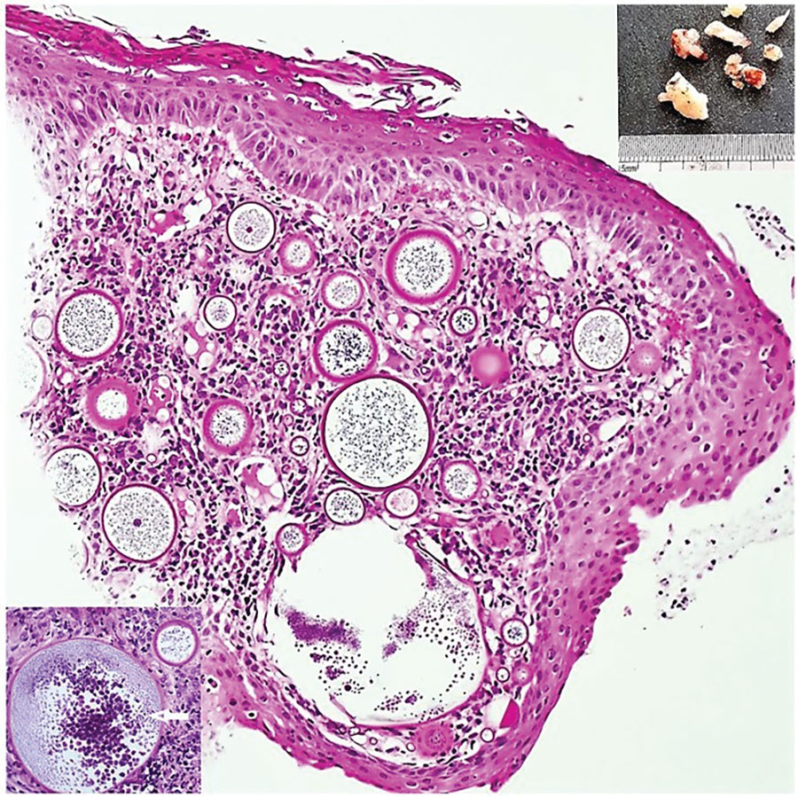
Figure 1. Results of testing in a 24-year-old Black woman with rhinosporidiosis, South Africa. Squamous mucosa with numerous thick-walled sporangia in the subepithelial region amid subacute inflammation. Hematoxylin and eosin stained section; original magnification ×100. Upper right inset shows polypoid solid fragments of tissue; lower left inset depicts sporangia enclosing endospores maturing centripetally (white arrow). Insets: original magnification ×200.
Page created: February 06, 2024
Page updated: March 20, 2024
Page reviewed: March 20, 2024
The conclusions, findings, and opinions expressed by authors contributing to this journal do not necessarily reflect the official position of the U.S. Department of Health and Human Services, the Public Health Service, the Centers for Disease Control and Prevention, or the authors' affiliated institutions. Use of trade names is for identification only and does not imply endorsement by any of the groups named above.