Volume 30, Number 4—April 2024
Dispatch
Case Report of Nasal Rhinosporidiosis in South Africa
Huzaifah Mayet, Denasha L. Reddy, Tika Bello Alvarez, Yahya Atiya, Nelesh P. Govender, Monica Birkhead , Tsidiso Maphanga, and Sugeshnee Pather
Figure 2
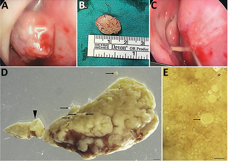
Figure 2. Macromorphology of excised recurrent nasal polyp from a 24-year-old Black woman with rhinosporidiosis, South Africa. A) Intraoperative endoscopic image of mass in right nasal cavity. B) Polypoid, oval mass measuring 15 mm. C) Stalk that attached the mass to the nasal septum. D) Portion of the pedunculated polyp (arrowhead) dotted with developing and mature sporangia (arrows). Scale bar = 1 mm. E) Surface of tissue with multiple sporangia in various stages of maturity, with the chitinous wall thickening during maturation (arrow). Scale bar = 150 μm.
Page created: February 06, 2024
Page updated: March 20, 2024
Page reviewed: March 20, 2024
The conclusions, findings, and opinions expressed by authors contributing to this journal do not necessarily reflect the official position of the U.S. Department of Health and Human Services, the Public Health Service, the Centers for Disease Control and Prevention, or the authors' affiliated institutions. Use of trade names is for identification only and does not imply endorsement by any of the groups named above.