Volume 30, Number 5—May 2024
Research Letter
Novel Patterns in High-Resolution Computed Tomography in Whipple Pneumonia
Figure
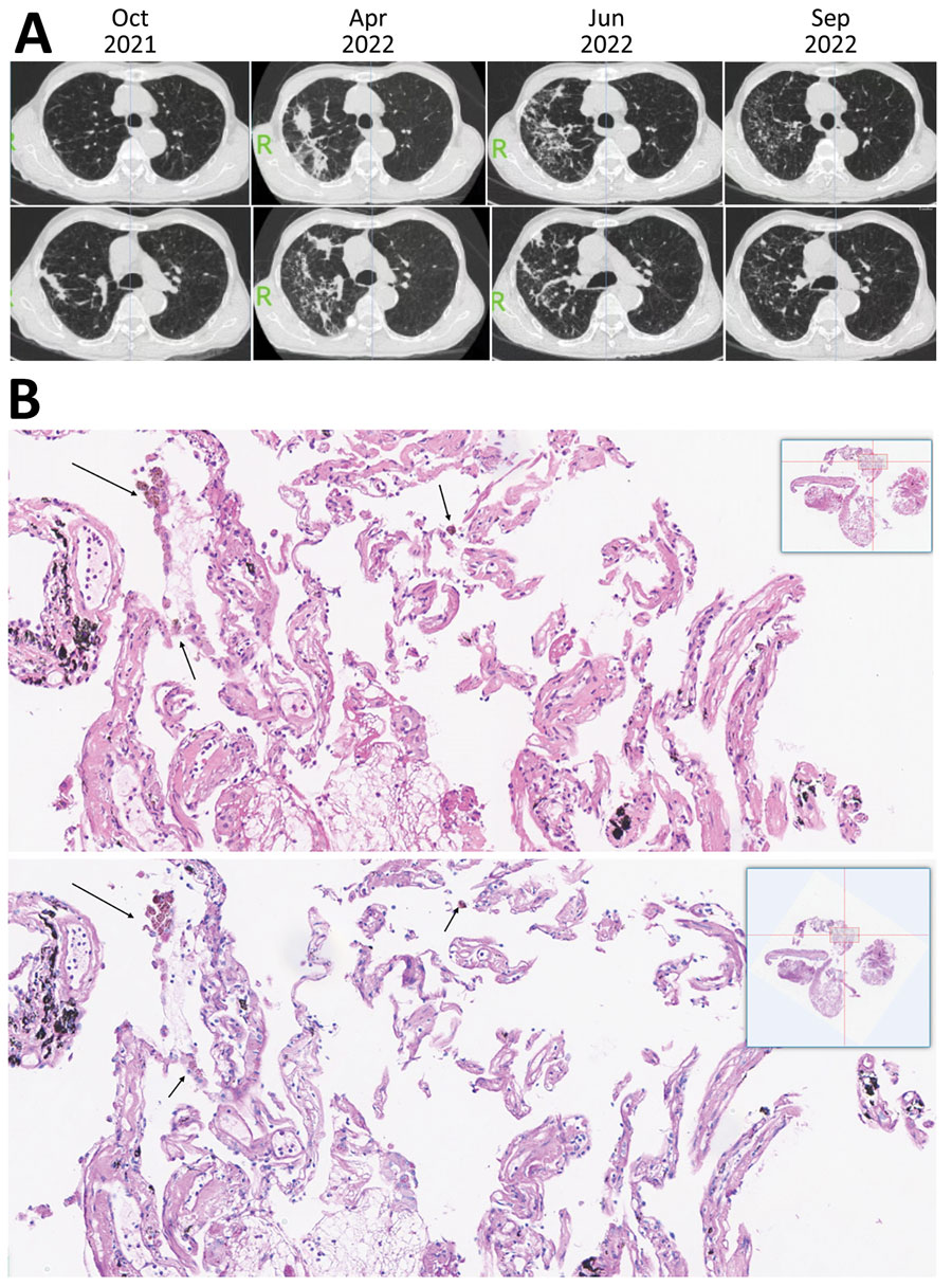
Figure. High-resolution computed tomography imaging and histology findings of the lung biopsy from a 67-year-old patient in China who had Tropheryma whipplei pneumonia. A) High-resolution computed tomography imaging showing gradual increase of diffused micronodules gathering in the upper right lung 6 months before diagnosis. In October 2021, micronodular and cord-like consolidation were seen on the upper right lung. In April 2022, the lesions were seen changing on both range and pattern and forming movable properties. In June 2022, the lesions were changed and scattered compared with lesions observed in April 2022. In September 2022, lesions were absorbed after 3 months of combined therapy consisting of minocycline and hydroxychloroquine. B) Magnified portion of slide showing histologic findings from the lung biopsy of the patient. The top image shows increased foamy macrophages within alveolar space, thickened alveolar septal and collagen deposition. The top image stain is hematoxylin and eosin staining, with arrows indicating foamy macrophages that have phagocytosed carbon pigment; the bottom image is periodic acid-Schiff staining and is negative for foamy macrophages. Insets show the entire histology slide.