Skip directly to site content Skip directly to page options Skip directly to A-Z link Skip directly to A-Z link Skip directly to A-Z link
Volume 30, Number 6—June 2024
Dispatch

Choanephora infundibulifera Rhinosinusitis in Man with Acute Lymphoblastic Leukemia, Tennessee, USA

Anita Max, Heather L. Glasgow, Teresa C.B. Santiago, Ashley Holland, Hiroto Inaba, Connie F. Cañete-Gibas, Nathan P. Wiederhold, Randall T. Hayden, and Elisabeth E. AddersonComments to Author 
Author affiliations: St. Jude Children’s Research Hospital, Memphis, Tennessee, USA (A. Max, H.L. Glasgow, T.C.B. Santiago, A. Holland, H. Inaba, E.E. Adderson); University of Tennessee Health Sciences Center, Memphis (H. Inaba, E.E. Adderson); University of Texas Health Science Center, San Antonio, Texas, USA (C.F. Cañete-Gibas, N.P. Wiederhold)

Main Article

Figure 1

Computed tomography, endoscopic findings, and histomorphology of rhinosinusitis caused by Choanephora infundibulifera in a man with leukemia in Memphis, Tennessee, USA. A) Computed tomography shows new asymmetric swelling of the anterior nasal septum and irregularity of the right septum, edema of the inferior turbinates, and obstruction of the right frontal sinus outflow tract. A septal perforation, the sequela of the patient’s previous fungal rhinosinusitis, was stable. B) Nasal endoscopy reveals necrosis of the anterior nasal septa. C, D) Necrotic sinonasal mucosa contains numerous hyaline (nonpigmented) fungal elements with broad (ribbon-like), thin-walled, nonseptated, and pleomorphic fungal hyphae. Hematoxylin and eosin stain; original magnification ×200 for panel C, ×400 for panel D. E, F) Gomori methenamine-silver stain highlights the fungal elements (in black). Original magnification ×200 for panel E, ×400 for panel F.

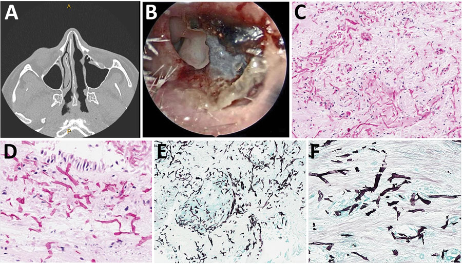
Figure 1. Computed tomography, endoscopic findings, and histomorphology of rhinosinusitis caused by Choanephora infundibulifera in a man with leukemia in Memphis, Tennessee, USA. A) Computed tomography shows new asymmetric swelling of the anterior nasal septum and irregularity of the right septum, edema of the inferior turbinates, and obstruction of the right frontal sinus outflow tract. A septal perforation, the sequela of the patient’s previous fungal rhinosinusitis, was stable. B) Nasal endoscopy reveals necrosis of the anterior nasal septa. C, D) Necrotic sinonasal mucosa contains numerous hyaline (nonpigmented) fungal elements with broad (ribbon-like), thin-walled, nonseptated, and pleomorphic fungal hyphae. Hematoxylin and eosin stain; original magnification ×200 for panel C, ×400 for panel D. E, F) Gomori methenamine-silver stain highlights the fungal elements (in black). Original magnification ×200 for panel E, ×400 for panel F.

Main Article

Page created: May 02, 2024
Page updated: May 22, 2024
Page reviewed: May 22, 2024
The conclusions, findings, and opinions expressed by authors contributing to this journal do not necessarily reflect the official position of the U.S. Department of Health and Human Services, the Public Health Service, the Centers for Disease Control and Prevention, or the authors' affiliated institutions. Use of trade names is for identification only and does not imply endorsement by any of the groups named above.
file_external