Skip directly to site content Skip directly to page options Skip directly to A-Z link Skip directly to A-Z link Skip directly to A-Z link
Volume 30, Number 6—June 2024
Research

Outbreak of Highly Pathogenic Avian Influenza A(H5N1) Virus in Seals, St. Lawrence Estuary, Quebec, Canada1

Stéphane LairComments to Author , Louise Quesnel, Anthony V. Signore, Pauline Delnatte, Carissa Embury-Hyatt, Marie-Soleil Nadeau, Oliver Lung, Shannon T. Ferrell, Robert Michaud, and Yohannes Berhane
Author affiliations: Université de Montréal, St. Hyacinthe, Quebec, Canada (S. Lair, L. Quesnel, P. Delnatte, S.T. Ferrell); Canadian Food Inspection Agency, Winnipeg, Manitoba, Canada (A.V. Signore, C. Embury-Hyatt, O. Lung, Y. Berhane); Ministère de l’Agriculture, des Pêcheries et de l’Alimentation du Quebec, St. Hyacinthe (M.-S. Nadeau); Groupe de recherche et d’éducation sur les mammifères marins, Tadoussac, Quebec, Canada (R. Michaud).

Main Article

Figure 3

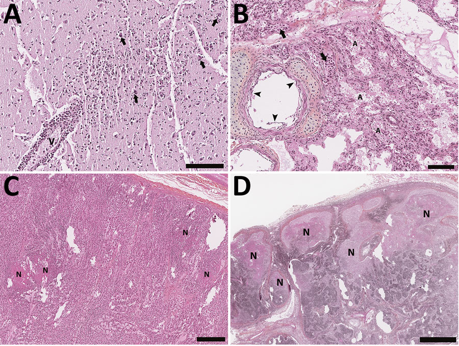
Histology section of thawed, formalin-fixed tissues from harbor seals (Phoca vitulina) infected by highly pathogenic avian influenza A(H5N1) virus in the St. Lawrence Estuary, Quebec, Canada, 2022. Hematoxylin phloxine saffron stain. A) Brain tissue from a young (<1 year old) female seal. The Virchow-Robin space around a vessel (V) is infiltrated by numerous layers of polymorphonuclear cells. Several neurons have a condensed hyperacidophilic cytoplasm indicative of necrosis and are often associated with satellitosis (arrows). A focally extensive infiltration of the neuropil by neutrophils and glial cells is also present. Scale bar indicates 100 µm. B) Lung from a young (<1 year old) female seal. The alveolar (A) and vascular (arrow) lumens contain numerous, often degenerate, polymorphonuclear cells. The alveolar walls are infiltrated by numerous inflammatory cells composed of neutrophils and mononuclear cells. The epithelial cells bordering the small bronchi are often necrotic. Scale bar indicates 100 µm. C) Adrenal gland of an adult female seal. Multifocal foci of necrosis are present in the cortical zone (N). Scale bar indicates 300 µm. D) Lymph node from an adult female seal. Marked multifocal to coalescing necrosis of the lymphoid tissues in the cortical region are noted (N). Scale bar indicates 1 mm.

Figure 3. Histology section of thawed, formalin-fixed tissues from harbor seals (Phoca vitulina) infected by highly pathogenic avian influenza A(H5N1) virus in the St. Lawrence Estuary, Quebec, Canada, 2022. Hematoxylin phloxine saffron stain. A) Brain tissue from a young (<1 year old) female seal. The Virchow-Robin space around a vessel (V) is infiltrated by numerous layers of polymorphonuclear cells. Several neurons have a condensed hyperacidophilic cytoplasm indicative of necrosis and are often associated with satellitosis (arrows). A focally extensive infiltration of the neuropil by neutrophils and glial cells is also present. Scale bar indicates 100 µm. B) Lung from a young (<1 year old) female seal. The alveolar (A) and vascular (arrow) lumens contain numerous, often degenerate, polymorphonuclear cells. The alveolar walls are infiltrated by numerous inflammatory cells composed of neutrophils and mononuclear cells. The epithelial cells bordering the small bronchi are often necrotic. Scale bar indicates 100 µm. C) Adrenal gland of an adult female seal. Multifocal foci of necrosis are present in the cortical zone (N). Scale bar indicates 300 µm. D) Lymph node from an adult female seal. Marked multifocal to coalescing necrosis of the lymphoid tissues in the cortical region are noted (N). Scale bar indicates 1 mm.

Main Article

1Preliminary results from this study were presented at the International Association for Aquatic Animal Medicine Conference, May 24, 2023, Salt Lake City, Utah, USA.

Page created: April 23, 2024
Page updated: May 22, 2024
Page reviewed: May 22, 2024
The conclusions, findings, and opinions expressed by authors contributing to this journal do not necessarily reflect the official position of the U.S. Department of Health and Human Services, the Public Health Service, the Centers for Disease Control and Prevention, or the authors' affiliated institutions. Use of trade names is for identification only and does not imply endorsement by any of the groups named above.
file_external