Volume 30, Number 7—July 2024
Dispatch
Acute Meningoencephalitis Associated with Borrelia miyamotoi, Minnesota, USA
Jeffrey M. Kubiak , Michael Klevay, Evann E. Hilt, and Patricia Ferrieri
Figure
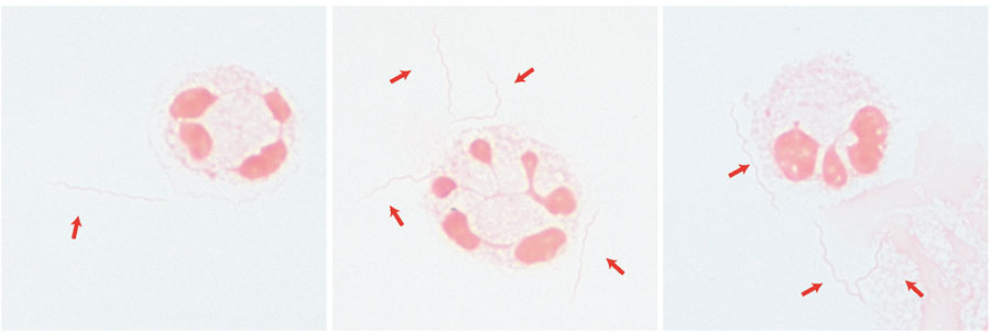
Figure. Spirochetes (red arrows) visualized on Gram stain in a cerebrospinal fluid sample from a 68-year-old man with immunosuppression from rituximab, Minnesota, USA. Visualization was done after concentration using cytospin (original magnification ×1,000 with oil immersion). The spirochetes were later identified as Borrelia miyamotoi by 16S ribosomal sequencing.
Page created: April 10, 2024
Page updated: June 22, 2024
Page reviewed: June 22, 2024
The conclusions, findings, and opinions expressed by authors contributing to this journal do not necessarily reflect the official position of the U.S. Department of Health and Human Services, the Public Health Service, the Centers for Disease Control and Prevention, or the authors' affiliated institutions. Use of trade names is for identification only and does not imply endorsement by any of the groups named above.