Volume 30, Number 7—July 2024
Dispatch
Acute Meningoencephalitis Associated with Borrelia miyamotoi, Minnesota, USA
Cite This Article
Citation for Media
Abstract
Borrelia miyamotoi is an emerging tickborne pathogen that has been associated with central nervous system infections in immunocompromised patients, albeit infrequently. We describe a case-patient in Minnesota, USA, who had meningeal symptoms of 1 month duration. B. miyamotoi infection was diagnosed by Gram staining on cerebrospinal fluid and confirmed by sequencing.
Borrelia miyamotoi is an emerging tickborne pathogen initially discovered in Japan in 1995 but not recognized as a human pathogen until described in a case series from Russia in 2011 (1). The bacterium has a geographic distribution across northern Europe and Asia, as well as in the northeastern and midwestern United States; the first case of B. miyamotoi in Minnesota, USA, was reported in 2016 (2). B. miyamotoi is spread by many species of the deer tick, including Ixodes scapularis in North America and I. ricinus in Europe and Asia. B. miyamotoi is classified within the relapsing-fever group of spirochetes; infection typically manifests as a nonspecific febrile illness with headaches, chills, myalgia, and arthralgia and is likely underdiagnosed because of its rarity and lack of specific symptoms (3,4).
B. miyamotoi has been associated with cases of neuroborreliosis in which spirochetes proliferate in the cerebrospinal fluid (CSF) to cause disease ranging from acute meningitis symptoms to prolonged encephalitis with altered mentation and cranial nerve deficits (5–8). Here, we report a case of acute meningoencephalitis associated with B. miyamotoi in the midwestern United States that was diagnosed on CSF Gram stain and confirmed by sequencing.
A 68-year-old man with immunosuppression from rituximab, in remission from mantle cell lymphoma involving the right eye, sought treatment at an emergency department in Minnesota in late summer. He had a 5-week history of headaches of increasing severity, worsened by positional changes and physical activity, as well as double vision, hearing difficulties, and some altered mentation. The patient was afebrile and had no nuchal rigidity. Cranial nerves exhibited no dysfunction other than subjective hearing loss, and physical examination results were otherwise unremarkable. He was an avid outdoorsman who had hiked frequently in the woodlands of Minnesota and Wisconsin over the months before seeking treatment, and he recalled identifying an engorged tick among several tick bites on his body during this time. He reported no travel history outside of the midwestern United States. The patient had been treated ≈1 year earlier with doxycycline for clinically diagnosed Lyme disease with fever and erythema migrans, but serologic B. burgdorferi testing at the time was negative.
We performed a magnetic resonance imaging scan of the brain and orbits with and without contrast, which showed bilateral abnormal cranial nerve enhancement of the oculomotor, trigeminal, facial, and vestibulocochlear nerves compared with a magnetic resonance image from 2 months earlier. Results of a lumbar puncture included a neutrophil-predominant pleocytosis (177 leukocytes/μL, 73% neutrophils). CSF flow cytometry results were unremarkable. A Biofire FilmArray (bioMérieux, https://www.biomerieux.com) meningitis/encephalitis multiplex PCR panel was negative. The patient had an elevated C-reactive protein (19.5 mg/L). Serologic results for B. burgdorferi in both the blood and CSF samples were negative, as were results of antitreponemal testing. Blood culture results were negative, and routine laboratory values were otherwise unremarkable.
Figure
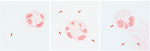
Figure. Spirochetes (red arrows) visualized on Gram stain in a cerebrospinal fluid sample from a 68-year-old man with immunosuppression from rituximab, Minnesota, USA. Visualization was done after concentration using cytospin (original...
A direct CSF Gram stain was performed in the University of Minnesota Medical Center Infectious Diseases Diagnostic Laboratory (Minneapolis, MN, USA) (Figure). The CSF specimen showed numerous gram-negative bacteria that measured 10–20 μm long with a tight coil, consistent with spirochetes. The CSF specimen was sent to the University of Washington Molecular Diagnosis Microbiology Section (Seattle, WA, USA) for bacterial 16S ribosomal RNA (rRNA) sequencing and fungal 28S rRNA sequencing.
The patient was admitted and treated with 2 g intravenous ceftriaxone daily for presumed neuroborreliosis, pending identification of the organism. After administering the initial antimicrobial treatment, we sent a plasma sample for a tickborne relapsing fever PCR panel, which broadly detects Borrelia species including B. hermsii, B. parkeri, B. turicatae, and B. miyamotoi; the test result was negative. Within 24 hours, the patient’s condition improved on antimicrobial therapy, and on day 3 of hospitalization, we discharged him; he reported no headaches and some improvement in his cognition, hearing, and vision. Six days after we discharged the patient from the hospital, his 16S ribosomal sequencing result returned positive results for B. miyamotoi. We administered daily outpatient infusion therapy with ceftriaxone for 4 weeks, during which time he reported mild residual subjective memory loss, hearing loss, and loss of visual acuity from baseline, although double vision resolved. We advised him on appropriate tick-prevention methods because his history of outdoor activity indicated increased risk for future tick bites.
In cases of B. miyamotoi with neurologic manifestations, clinical symptoms have ranged from acute meningeal symptoms, including headache, vomiting, and dizziness (5,9), to prolonged neurologic disease, in some cases involving several months of progressive mental decline, confusion, gait instability, and hearing difficulty (6,7). Typically, in acute meningoencephalitis cases, spirochetes cannot be visualized in direct CSF specimen Gram or Giemsa stains (5). Diagnosis in those cases has relied on either broad-based 16S sequencing of bacteria rRNA, targeted PCR identification of the GlpQ or flagellin genes, darkfield microscopy, or acridine orange stain (5,6,9,10). In contrast, in 1 case, spirochetes were visualized on direct specimen Giemsa and Gram stains in the presence of prolonged neuroborreliosis of ≈4 months duration (7). Although literature on the subject remains lacking because of the relative rarity of this disease manifestation, those cases suggest that, in immunocompromised patients, spirochetes may proliferate in CSF over time, leading to an increased likelihood of detecting spirochetes on direct specimen examination in patients with prolonged symptoms. It is notable that DNA from B. miyamotoi was not detected in our patient’s plasma, suggesting a neurologic tropism that enabled continued spirochetal proliferation even after the organisms were cleared from the patient’s bloodstream.
This patient had abnormal MRI findings, including abnormal cranial nerve enhancement of the oculomotor, trigeminal, facial, and vestibulocochlear cranial nerves. Those findings were initially worrisome for leptomeningeal progression of the patient’s ocular lymphoma, but flow cytometry of his CSF did not demonstrate abnormal proliferation of B cells. Although previous case reports have shown no abnormalities on cranial MRI (5–7), given this patient’s constellation of symptoms, we believe that these MRI findings were attributable to this infection and the proliferation of spirochetes in the CSF.
Differential diagnosis of spirochetes visualized on CSF Gram stain is limited. We found 1 other published case report with this finding, also for B. miyamotoi (7). Although other spirochetes, such as B. burgdorferi and Treponema pallidum, are known to cause neurologic manifestations ranging from acute meningoencephalitis to chronic encephalitis, those organisms are not visualized with Gram stain; diagnoses require darkfield microscopy, PCR, or serologic evidence of disease (10–13). We posit that finding spirochetes on CSF Gram stain is highly suggestive of B. miyamotoi, and patients should be empirically treated for neuroborreliosis pending definitive identification, particularly patients active in geographic areas with known tick exposure who have rituximab-caused immunosuppression.
Of note, most reported cases of neuroborreliosis caused by B. miyamotoi have been found in older patients on rituximab therapy with B-cell depletion, as in this case-patient, although there are rare reports of neuroborreliosis in immunocompetent persons (9). The adaptive immune system is considered key in controlling B. miyamotoi infection; B. miyamotoi activates dendritic cells, phagocytizing the bacteria and stimulating release of several cytokines (14). Furthermore, studies in related species, such as B. hermsii, have shown that clearance of spirochetes is mediated by antibodies (15). Further research into the pathogenesis of B. miyamotoi meningoencephalitis, with an emphasis on the regulators of proliferation of these organisms in CSF and disease progression in immunocompromised patients, are certainly warranted. Greater awareness of this manifestation of B. miyamotoi disease is needed to increased diagnostic accuracy and antimicrobial treatment to enable improved patient outcomes.
Dr. Kubiak is the assistant medical director of the Infectious Diseases Diagnostic Laboratory at the M Health Fairview University of Minnesota Medical Center, where he is an assistant professor in the Department of Laboratory Medicine and Pathology. His primary research interests are in new laboratory diagnostics and laboratory informatics.
Acknowledgment
We thank the patient for agreeing to the publication of this report. In addition, we thank the technical staff of the Infectious Diseases Diagnostic Laboratory for considering the spirochetal possibility on the CSF direct Gram stain.
References
- Platonov AE, Karan LS, Kolyasnikova NM, Makhneva NA, Toporkova MG, Maleev VV, et al. Humans infected with relapsing fever spirochete Borrelia miyamotoi, Russia. Emerg Infect Dis. 2011;17:1816–23. DOIPubMedGoogle Scholar
- Minnesota Department of Health. About Borrelia miyamotoi disease [cited 2023 Nov 21]. https://www.health.state.mn.us/diseases/bmiyamotoi/basics.html
- Cleveland DW, Anderson CC, Brissette CA. Borrelia miyamotoi: a comprehensive review. Pathogens. 2023;12:267. DOIPubMedGoogle Scholar
- McCormick DW, Brown CM, Bjork J, Cervantes K, Esponda-Morrison B, Garrett J, et al. Characteristics of hard tick relapsing fever caused by Borrelia miyamotoi, United States, 2013–2019. Emerg Infect Dis. 2023;29:1719–29. DOIPubMedGoogle Scholar
- Boden K, Lobenstein S, Hermann B, Margos G, Fingerle V. Borrelia miyamotoi–associated neuroborreliosis in immunocompromised person. Emerg Infect Dis. 2016;22:1617–20. DOIPubMedGoogle Scholar
- Hovius JWR, de Wever B, Sohne M, Brouwer MC, Coumou J, Wagemakers A, et al. A case of meningoencephalitis by the relapsing fever spirochaete Borrelia miyamotoi in Europe. Lancet. 2013;382:658. DOIPubMedGoogle Scholar
- Gugliotta JL, Goethert HK, Berardi VP, Telford SR III. Meningoencephalitis from Borrelia miyamotoi in an immunocompromised patient. N Engl J Med. 2013;368:240–5. DOIPubMedGoogle Scholar
- Kubiak K, Szczotko M, Dmitryjuk M. Borrelia miyamotoi—an emerging human tick-borne pathogen in Europe. Microorganisms. 2021;9:154. DOIPubMedGoogle Scholar
- Henningsson AJ, Asgeirsson H, Hammas B, Karlsson E, Parke Å, Hoornstra D, et al. Two cases of Borrelia miyamotoi meningitis, Sweden, 2018. Emerg Infect Dis. 2019;25:1965–8. DOIPubMedGoogle Scholar
- Gyllemark P, Wilhelmsson P, Elm C, Hoornstra D, Hovius JW, Johansson M, et al. Are other tick-borne infections overlooked in patients investigated for Lyme neuroborreliosis? A large retrospective study from South-eastern Sweden. Ticks Tick Borne Dis. 2021;12:
101759 . DOIPubMedGoogle Scholar - Madison-Antenucci S, Kramer LD, Gebhardt LL, Kauffman E. Emerging tick-borne diseases. Clin Microbiol Rev. 2020;33:e00083–18. DOIPubMedGoogle Scholar
- Ramchandani MS, Cannon CA, Marra CM. Syphilis: a modern resurgence. Infect Dis Clin North Am. 2023;37:195–222. DOIPubMedGoogle Scholar
- Mason LMK, Koetsveld J, Trentelman JJA, Kaptein TM, Hoornstra D, Wagemakers A, et al. Borrelia miyamotoi activates human dendritic cells and elicits T cell responses. J Immunol. 2020;204:386–93. DOIPubMedGoogle Scholar
- Crowder CD, Ghalyanchi Langeroudi A, Shojaee Estabragh A, Lewis ERG, Marcsisin RA, Barbour AG. Pathogen and host response dynamics in a mouse model of Borrelia hermsii relapsing fever. Vet Sci. 2016;3:19. DOIPubMedGoogle Scholar
Figure
Cite This ArticleOriginal Publication Date: June 18, 2024
Table of Contents – Volume 30, Number 7—July 2024
| EID Search Options |
|---|
|
|
|
|
|
|
Please use the form below to submit correspondence to the authors or contact them at the following address:
Jeffrey M. Kubiak, Department of Laboratory Medicine and Pathology, University of Minnesota Medical Center, 420 Delaware St SE, Minneapolis, MN 55455, USA
Top