Volume 30, Number 8—August 2024
Research Letter
Rare Case of Echinostoma cinetorchis Infection, South Korea
Figure 1

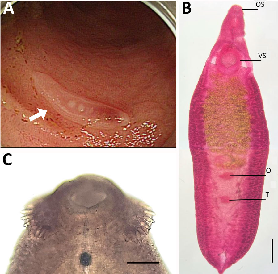
Figure 1. Analysis of a worm identified as Echinostoma cinetorchis removed during colonoscopy from a 69-year-old woman in South Korea. A) Colonoscopy image showing a moving trematode in the mucosa of the descending colon. B) Whole body of the worm. Scale bar = 0.6 mm. C) Head part of the worm showing collar spines (37 in total number) on the head collar around the oral sucker, by which it could be morphologically identified as a 37-collar-spined echinostome. Scale bar = 0.1 mm). O, ovary; OS, oral sucker; T, testis; VS, ventral sucker.
Page created: June 28, 2024
Page updated: July 22, 2024
Page reviewed: July 22, 2024
The conclusions, findings, and opinions expressed by authors contributing to this journal do not necessarily reflect the official position of the U.S. Department of Health and Human Services, the Public Health Service, the Centers for Disease Control and Prevention, or the authors' affiliated institutions. Use of trade names is for identification only and does not imply endorsement by any of the groups named above.