Volume 30, Number 9—September 2024
Research Letter
Confirmed Case of Autochthonous Human Babesiosis, Hungary
Dávid Sipos , Ágnes Kappéter, Barbara Réger, Gabriella Kiss, Nóra Takács, Róbert Farkas, István Kucsera, and Zoltán Péterfi
Figure 1
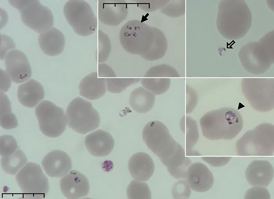
Figure 1. Peripheral blood smear from patient who had a confirmed case of autochthonous human babesiosis, Hungary. Smear shows erythrocytes infected with Babesia sp.; smear was stained with May-Grünwald Giemsa stain and examined by using light microscopy. In inset images, solid arrow indicates cells infected with multiple merozoites, open arrow indicates extracellular parasites, and arrowhead indicates vacuolated ring forms (trophozoites). Scale bars indicates 10 μm.
Page created: July 23, 2024
Page updated: August 21, 2024
Page reviewed: August 21, 2024
The conclusions, findings, and opinions expressed by authors contributing to this journal do not necessarily reflect the official position of the U.S. Department of Health and Human Services, the Public Health Service, the Centers for Disease Control and Prevention, or the authors' affiliated institutions. Use of trade names is for identification only and does not imply endorsement by any of the groups named above.