Endogenous Endophthalmitis Caused by Prototheca Microalga in Birman Cat, Spain
Laura Jimenez-Ramos
1, Ana Ripolles-Garcia
1, Gianvito Lanave, Francesco Pellegrini, Miriam Caro-Suarez, Almudena Latre-Moreno, Marta Ferruz-Fernandez, Maria Luisa Palmero-Colado, Vanessa Carballes-Perez, Antonio Melendez-Lazo, Carolina Naranjo, Fernando Laguna

, Vito Martella, and Manuel Villagrasa
Author affiliation: Puchol Veterinary Hospital, Madrid, Spain (L. Jimenez-Ramos, A. Ripolles-Garcia, M. Caro-Suarez, A. Latre-Moreno, M. Ferruz-Fernandez, F. Laguna, M. Villagrasa); Centro Oftalmológico Veterinario Goya, Madrid (L. Jimenez-Ramos, A. Ripolles-Garcia, M. Caro-Suarez, A. Latre-Moreno, M. Ferruz-Fernandez, F. Laguna, M. Villagrasa); University of Bari Aldo Moro, Bari, Italy (G. Lanave, F. Pellegrini, V. Martella); Gattos Veterinary Hospital, Madrid (M.L. Luisa Palmero-Colado, V. Carballes-Perez); T-Cito Laboratories, Barcelona, Spain (A. Melendez-Lazo); IDEXX Laboratories, Barcelona (C. Naranjo); University of Veterinary Medicine, Budapest, Hungary (V. Martella)
Main Article
Figure

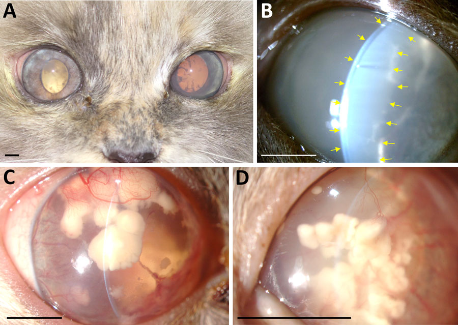
Figure. Clinical course of bilateral endogenous endophthalmitis in 5-year-old female Birman cat evaluated by slit-lamp biomicroscopic examination, Madrid, Spain. A) Digital photograph of both eyes demonstrating bilateral uveitis at 5.5 months after initial visit to clinic. B) Slit-lamp biomicroscopic image (original magnification ×10) of the left eye, demonstrating a marked flare (yellow arrows) at 6.5 months after initial clinical signs. C, D) At 16.5 months, the right eye (C) (original magnification ×10) and left eye (D) (original magnification ×16) were imaged by slit-lamp biomicroscopic examination, revealing corneal endothelial macrodeposits of undefined origin, presumed to be the result of Prototheca spp. invasion. Scale bars indicate 5 mm.
Main Article
Page created: December 06, 2024
Page updated: December 22, 2024
Page reviewed: December 22, 2024
The conclusions, findings, and opinions expressed by authors contributing to this journal do not necessarily reflect the official position of the U.S. Department of Health and Human Services, the Public Health Service, the Centers for Disease Control and Prevention, or the authors' affiliated institutions. Use of trade names is for identification only and does not imply endorsement by any of the groups named above.
