Volume 31, Number 10—October 2025
Synopsis
Organ Donor Transmission of Rickettsia typhi to Kidney Transplant Recipients, Texas, USA, 2024
Jeffrey C. Jones, Omar G. García, Julian A. Villalba, Rosa Hinojosa, Marissa L. Taylor, Pallavi Annambhotla, Matthias H. Kapturczak, Bonny Mayes, Sandor E. Karpathy, Arlyn N. Gleaton, Linda Moon, Joseph Singleton, Sridhar V. Basavaraju, and Christopher D. Paddock
Figure 2
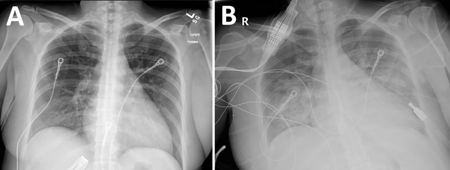
Figure 2. Chest radiographs from an organ donor with murine typhus, from a study describing the transmission of Rickettsia typhi from an organ donor to 2 kidney transplant recipients, Texas, 2024. A) Chest radiograph obtained at hospital admission, 5 days after symptom onset, with no specific abnormalities noted. B) Chest radiograph obtained on hospital day 3, revealing extensive alveolar opacities predominantly in the middle and lower lung fields.
Page created: September 09, 2025
Page updated: September 25, 2025
Page reviewed: September 25, 2025
The conclusions, findings, and opinions expressed by authors contributing to this journal do not necessarily reflect the official position of the U.S. Department of Health and Human Services, the Public Health Service, the Centers for Disease Control and Prevention, or the authors' affiliated institutions. Use of trade names is for identification only and does not imply endorsement by any of the groups named above.