Skip directly to site content Skip directly to page options Skip directly to A-Z link Skip directly to A-Z link Skip directly to A-Z link

Volume 31, Number 10—October 2025

Synopsis

Organ Donor Transmission of Rickettsia typhi to Kidney Transplant Recipients, Texas, USA, 2024

Jeffrey C. Jones, Omar G. García, Julian A. Villalba, Rosa Hinojosa, Marissa L. Taylor, Pallavi Annambhotla, Matthias H. Kapturczak, Bonny Mayes, Sandor E. Karpathy, Arlyn N. Gleaton, Linda Moon, Joseph Singleton, Sridhar V. Basavaraju, and Christopher D. PaddockComments to Author 
Author affiliation: San Antonio Infectious Diseases Consultants, San Antonio, Texas, USA (J.C. Jones); City of Laredo Public Health Department, Laredo, Texas, USA (O.G. García); Centers for Disease Control and Prevention, Atlanta, Georgia, USA (J.A. Villalba, M.L. Taylor, P. Annambhotla, S.E. Karpathy, A.N. Gleaton, L. Moon, J. Singleton, S.V. Basavaraju, C.D. Paddock); Corpus Christi Nueces County Public Health District, Corpus Christi, Texas, USA (R. Hinojosa); Methodist Transplant Institute, San Antonio (M.H. Kapturczak); Texas Department of State Health Services, Austin, Texas, USA (B. Mayes).

Main Article

Figure 3

Histopathological and immunohistochemical features of formalin-fixed, paraffin-embedded biopsy specimens collected from the right and left kidney allografts procured from a donor who died of murine typhus, from a study describing the transmission of Rickettsia typhi from an organ donor to 2 kidney transplant recipients, Texas, 2024. A) When stained with hematoxylin and eosin, both allografts showed multifocal, interstitial, and predominantly mononuclear peritubular infiltrates, associated with focal endarteritis (thin arrows), features of acute tubular injury, including epithelial attenuation with loss of apical cytoplasm, and pigmented casts (large arrow). B) Glomeruli displayed moderate mesangial hypercellularity and tuft adhesions to the Bowman’s capsules (arrows) and intracapillary and mesangial phagocytic foam-cells. C) Short chains comprising small, rod-shaped bacteria were revealed in glomerular capillaries by the Warthin–Starry silver impregnation staining technique. D–F) An immunohistochemical stain for typhus-group rickettsiae revealed intact bacteria within endothelial cells of inflamed small vessels and vascular spaces of mesangial capillaries (arrows) and phagocytized bacterial antigens in the cytoplasm of glomerular foam-cells (arrows). Original magnifications ×400 (A, B, and E) and ×1,000 (C, D, and F).

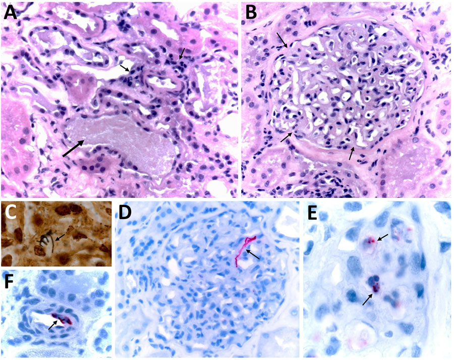
Figure 3. Histopathological and immunohistochemical features of formalin-fixed, paraffin-embedded biopsy specimens collected from the right and left kidney allografts procured from a donor who died of murine typhus, from a study describing the transmission of Rickettsia typhi from an organ donor to 2 kidney transplant recipients, Texas, 2024. A) When stained with hematoxylin and eosin, both allografts showed multifocal, interstitial, and predominantly mononuclear peritubular infiltrates, associated with focal endarteritis (thin arrows), features of acute tubular injury, including epithelial attenuation with loss of apical cytoplasm, and pigmented casts (large arrow). B) Glomeruli displayed moderate mesangial hypercellularity and tuft adhesions to the Bowman’s capsules (arrows) and intracapillary and mesangial phagocytic foam-cells. C) Short chains comprising small, rod-shaped bacteria were revealed in glomerular capillaries by the Warthin–Starry silver impregnation staining technique. D–F) An immunohistochemical stain for typhus-group rickettsiae revealed intact bacteria within endothelial cells of inflamed small vessels and vascular spaces of mesangial capillaries (arrows) and phagocytized bacterial antigens in the cytoplasm of glomerular foam-cells (arrows). Original magnifications ×400 (A, B, and E) and ×1,000 (C, D, and F).

Main Article

Page created: September 09, 2025
Page updated: September 25, 2025
Page reviewed: September 25, 2025
The conclusions, findings, and opinions expressed by authors contributing to this journal do not necessarily reflect the official position of the U.S. Department of Health and Human Services, the Public Health Service, the Centers for Disease Control and Prevention, or the authors' affiliated institutions. Use of trade names is for identification only and does not imply endorsement by any of the groups named above.
file_external