Volume 31, Number 11—November 2025
Synopsis
Trichosporon austroamericanum Infections among Hospitalized Patients, France, 2022–2024
Cite This Article
Citation for Media
Abstract
During 2022–2024, six cases of invasive fungal infection occurred among immunocompromised patients at Marseille University Hospital, Marseille, France. Matrix-assisted laser desorption/ionization time-of-flight mass spectrometry initially identified Trichosporon inkin fungi. However, phylogenetic analysis of intergenic spacer region 1 and whole-genome sequences revealed the genetically distinct species T. austroamericanum. Analysis of core genome and mitogenome from 6 patient isolates and 1 environmental isolate revealed substantial genetic diversity among T. austroamericanum strains, indicating a polyclonal outbreak. Furthermore, the mitochondrial genome emerged as a potential marker for intraspecies differentiation, which potentially could aid in epidemiologic investigations. Identified in 2024 but potentially underestimated, T. austroamericanum has since been reported in case clusters from hospital settings in France, highlighting the need for accurate fungal identification and suggesting previously identified T. inkin cases should be re-evaluated for T. austroamericanum. Clinical T. austroamericanum is emerging in hospital settings and should be included in the differential diagnosis of fungal infections.
Fungi belonging to various species of the genus Trichosporon can cause a wide range of infections, from superficial skin damage to serious systemic infections in immunocompromised persons, who can have high mortality rates (1). Trichosporon, which belongs to the Basidiomycota family, includes ≈20 species that are pathogenic to humans (2). Although Cutaneotrichosporon cutaneum and T. asahii are the predominant Basidiomycota species, new species have increasingly been described in human pathology since 2002 (2).
In 2024, a new Basidiomycota species, T. austroamericanum, was identified in a urine sample from a kidney transplant recipient in Brazil (3); the species was then detected in several clinical samples from France, South America, and Asia. T. austroamericanum and T. inkin have been confounded in the past before the species were distinguished through phylogenetic analysis of the intergenic spacer (IGS) 1 region and amplified fragment length polymorphism fingerprinting.
Ubiquitous in the environment, some Trichosporon species have been isolated from soil, leaf mold, and decayed wood (4). The environmental reservoirs of T. inkin and T. austroamericanum remain unknown. Moreover, emergence of hospital trichosporonosis cases caused by T. asahii have been described (5), highlighting hospital sources during epidemiologic and environmental investigations. Thus, Trichosporon species should not be overlooked in the hospital environment.
During July 2022–June 2024, the kidney transplant department of Marseille University Hospital, Marseille, France, recorded 4 cases of invasive fungal infection. Whole-genome sequencing (WGS) was initially performed on the first 3 reported cases by using matrix-assisted laser desorption/ionization time-of-flight (MALDI-TOF) mass spectrometry, which identified the agent as T. inkin. Further investigation using other molecular methods revealed T. austroamericanum fungi as the causative agent. Because of the rarity of the species, we suspected a putative common source. Moreover, 2 other cases subsequently were diagnosed in patients admitted in cardiology and gastrointestinal surgery centers in 2 other hospitals of our institution. We investigated the sudden upsurge of trichosporonosis cases through WGS and phylogenetic analysis.
We prospectively identified patients over a 2-year period from 3 different sites of Marseille University Hospital system, referred to as hospitals A, B, and C. Our study included all patients with associated clinical signs of infection from whom T. austroamericanum fungi was isolated from sterile sites.
We collected retrospective medical data for each patient, when available, by using HOSPILINK DPI version 5.11.3P10.8.3 (Axigate Link, https://axigatelink.com) and processed data in an anonymized Excel 2013 file (Microsoft, https://www.microsoft.com). Data included demographic information, exposure factors, underlying diseases, and clinical signs and symptoms.
Inclusion and Ethics
We anonymized all identified T. austroamericanum strains recovered from clinical samples and stored them in Cryosysteme Protect bead tubes (Dutscher, https://www.dutscher.com) at −20°C. The hospital strain bank assigned a unique number to each sample: L0221, L0385, L0399, L0419, L0445, and L0458. The hospital routinely performed antifungal susceptibility testing by using the Sensititre YeastOne microdilution method (Thermo Fisher Scientific, https://www.thermofisher.com), according to the manufacturer’s instructions.
This study was reviewed and approved by the Ethics Committee of Assistance Publique Hôpitaux de Marseille (approval no. CSE24-49) and the Assistance-Publique-Hôpitaux de Marseille Health Data Access Portal (approval no. PADS24-275). Patients were informed of the research and their nonopposition to the use of their data was collected. In accordance with those committees and current regulations, written informed consent was not required. All potentially identifying information was removed in compliance with International Committee of Medical Journal Editors guidelines.
Mycologic Investigation
From identified patients, we collected cryptococcal antigen in serum by using Cryptococcal Antigen Lateral Flow Assay (IMMY, https://www.immy.com) and Platelia Aspergillus Antigen (Bio-Rad Laboratories, https://www.bio-rad.com). In addition, the hospital’s infection control team initiated an environmental investigation in 2023, which they continued in 2024 until the last infected patient was identified. The investigation focused on the rooms and units where the initial kidney transplant patients had been under care from the date of admission to discharge, including radiology, the intensive care unit (ICU), the urological surgery operating theater, and the dialysis unit. The team also investigated the nephrology unit, where patients were hospitalized for a week after kidney transplantation. The team collected air samples by using AESAP1075 Sampl’air Lite microbiological air sampler (AES Laboratories, https://www.chemeurope.com), collecting 330 liters on Sabouraud dextrose agar (SDA). The team collected water samples from faucets or showers in patients’ rooms in 250-mL bottles containing sodium thiosulfate. The team also collected surface samples by using eSwab Liquid Amies Elution Swabs (Copan, https://www.copangroup.com).
Next-Generation Sequencing
We performed DNA extraction on pure subculture from SDA supplemented with gentamicin and chloramphenicol (Bio-Rad), using previously described methods (6). We sequenced genomic DNA by using a paired-end strategy. We barcoded and prepared samples by using the COVIDseq Test sample prep kit (Illumina, https://www.illumina.com) adapted for fungi, in which the tagmentation step fragmented and tagged the DNA. We used limited cycle PCR amplification (12 cycles) to complete the tag adapters and introduce dual-index barcodes. After purification on ITB beads (Illumina), we normalized libraries to the same molarity, then pooled those into a single library for sequencing on the NovaSeq 6000 (Illumina). We loaded the pooled single-strand library onto the reagent cartridge and then onto the instrument along with the flow cell. We conducted automated cluster generation and paired-end sequencing of dual index reads in a single 25-hour run of 2 × 150 bp.
We also performed nanopore sequencing on individually sequenced genomic DNA by using the PromethION 2 Solo or GridION and the LSK109 Ligation Kit on a FLO-PRO002 flow cell (all Oxford Nanopore Technologies, https://nanoporetech.com). The end-prep step fixes specific nucleotides to use for adapter ligation, after which we purified the DNA on magnetic beads (CleanNA, https://www.cleanna.com). We activated the flow cell by adding a flush buffer and a tether from Flow Cell Priming Kit EXP-FLP002 (Oxford Nanopore Technologies). We then loaded the libraries onto the flow cell for a 72-hour run.
Bioinformatics
We converted Illumina binary base call (BCL) files into fastq files by using bclconverter version 4.2.4 (Illumina). We used Trimmomatic version 0.39 (7) to trim reads to a minimum Phred quality of 33 and minimum read length of 36 bp. We trimmed nanopore raw reads by using ProwlerTrimmer (8), a Phred quality of 20–35, and minimum read length of 1,000 bp.
We obtained complete genomes from mixed de novo assembly of Illumina and nanopore reads by using Unicycler version 0.4.4 (9). We removed contigs <1,000 bp in length. We determined GC content and evaluated contamination by using ContEst16S (EZBio https://www.ezbiocloud.net). We assessed assembly quality by using BUSCO version 5.7.1 and the fungi_odb10 database (10).
We used the Burrows-Wheeler aligner (Galaxy version 2.3 plus galaxy0; https://usegalaxy.eu) (12) to map filtered reads to the reference genome CBS 17435 (11) from the CBS culture collection (https://wi.knaw.nl/fungal_table) hosted by the Westerdijk Fungal Biodiversity Institute (Utrecht, the Netherlands). We generated consensus sequences by using iVar consensus (Galaxy version 1.4.4 plus galaxy0) (13), with depth coverage of >10 reads and base quality of Q20. We aligned the resulting sequences by using MAFFT (Galaxy version 7.526 plus galaxy2) (14).
We calculated pairwise single-nucleotide polymorphism (SNP) distances by using SNP distance matrix (Galaxy version 7.526 plus galaxy2) and identified SNPs by using the Find SNP Sites command (Galaxy version 2.5.1 plus galaxy0) (15). We aligned concatenated SNP lists and analyzed with IQ-TREE version 2.4.0 (http://www.iqtree.org) plus galaxy1 to generate a maximum-likelihood phylogenetic tree, which we visualized on iTOL (https://itol.embl.de). We calculated genome coverage with a custom Python script (Python Software Foundation, https://www.python.org).
We isolated mitochondrial genomes into a single circular contig ≈40-kb in size and aligned contigs by using mauve version 2.4.0 (16). We oriented contigs from the cytochrome c oxidase 1 start codon. When necessary, we generated reverse complements using an online tool (https://reverse-complement.com/terms.html), then annotated genomes by using GeSeq (https://chlorobox.mpimp-golm.mpg.de/geseq.html).
We mapped trimmed Illumina reads against the reference IGS1 sequence (CBS 17435) using Burrows-Wheeler aligner (12) and obtained consensus sequences by using sam2consensus (https://github.com/edgardomortiz/sam2consensus). We used MAFFT (https://mafft.cbrc.jp/alignment/server/index.html) (13) to align sequences and performed subsequent phylogenetic analysis by using IQ-TREE with 1,000 bootstraps (17).
Maximum-Likelihood Core-Genome Phylogenetic Analysis
We constructed a core-genome tree by using the Galaxy platform. We masked repeats with RepeatMasker (Galaxy version 4.1.5 plus galaxy0) and annotated the assemblies by using the Maker annotation pipeline (18) with the Augustus predefined prediction model Cryptococcus neoformans gattii (Galaxy version 2.31.11 plus galaxy2). We derived the aligned core-genome from the resulting annotated general feature format files by using Roary in Galaxy version 3.13.0 plus galaxy3, then collected SNPs by using the Find SNP Sites tool (Galaxy Version 2.5.1 plus galaxy0). We used those SNPs to build the maximum-likelihood tree on IQ-TREE version 2.3.6 (built August 4, 2024) (17), under ModelFinder (19). We visualized trees by using the iTOL platform.
Patient Data
By June 2024, a total of 6 patients with T. austroamericanum infection had been identified (Table 1). The patients’ median age was 65.5 (range 55–83) years; 3 (50%) were male and 3 (50%) were female. Four patients were kidney transplant recipients who had clinical signs of an infection 2–4 months after transplantation; all 4 had scar dehiscence and subcutaneous abscesses, suggesting that the scar was the portal of entry.
The 2 other patients were immunocompetent. One (sample no. L0419) had been hospitalized in the cardiology department and was referred for a nephrology consultation during the time the outbreak was occurring. That patient had not undergone recent surgery. The other patient (sample no. L0445) experienced shock and acute respiratory distress syndrome after gastrointestinal surgery.
Of the 25 T. austroamericanum–positive samples, 16 (64%) were blood cultures from 2 patients, 3 were scar swab samples from 1 patient, 3 were abscess aspiration samples from 3 patients, and 3 were respiratory samples (2 bronchial aspirate and 1 bronchoalveolar lavage fluid) from 1 patient. All T. austroamericanum isolates exhibited similar antifungal susceptibility profiles (Table 2), and we noted no significant difference related to date of isolate collection.
Mycological Investigation
The infection control team conducted an environmental survey in hospital A during April 20, 2023–July 19, 2024. In total, they collected 145 surface samples, 33 air samples, and 17 water samples in the urological surgery operating theater and the following patient units: the nephrology ICU, the kidney transplantation unit, the nephrology daycare, the radiology unit, and the dialysis unit. Samples were mainly collected from patient rooms, medical device storage area, and the decontamination room. All 195 samples were negative for T. austroamericanum. However, a T. austroamericanum strain was isolated from an air sample taken as part of a routine surveillance in a pediatric ICU of hospital B.
Next-Generation Sequencing
Multilocus Sequence Typing
Figure 1
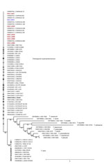
Figure 1. Maximum-likelihood phylogenetic tree of IGS1 sequences from study of Trichosporon austroamericanuminfections among hospitalized patients, France, 2022–2024. The tree includes strains isolated from 6 patients (red font) and 1...
We initially performed WGS on the first 3 reported cases, which MALDI-TOF mass spectrometry had identified as T. inkin. To investigate genetic variations, we applied a multilocus sequence typing approach. We mapped sequences of cytb, rpb1/2, and tef1 genes and the SSU and D1/D2 regions against the reference genome of T. inkin (GenBank accession no. MT801082). Although the sequences were identical among the isolates, notable differences from T. inkin emerged among isolates from those 3 cases. In addition, we observed high divergence between the strains from our investigation and the T. inkin reference mitochondrial genome and IGS1 marker, characterized by numerous point mutations, deletions, and insertions (Appendix Figure 1). The consistent mutation patterns across the strains, along with their divergence from T. inkin, strongly supported the classification of the isolates as a distinct taxonomic entity. That preliminary analysis served as the starting point for further investigation into the IGS1 region of the 6 patient-derived isolates. Phylogenetic analysis, which included 3 reference sequences provided by the National Reference Center for Invasive Mycoses and Antifungals (CNRMA) at Institut Pasteur (https://www.pasteur.fr), confirmed that the 7 strains (6 patient and 1 environmental) belonged to T. austroamericanum species (Figure 1).
WGS Typing
We conducted WGS typing to assess the clonal nature of the T. austroamericanum outbreak. Of the 7 genomes, 6 demonstrated excellent assembly quality and had contigs ranging from 12 to 31 (Table 3). Those assemblies showed consistent genome lengths of 20.8 to 21 Mb, and GC content was close to 61.31%. The BUSCO scores were all >96% (range 96.5%–97.4%), indicating highly complete and accurate assemblies. However, the assembly for strain L0445 stood out because it had a much higher number of contigs (1,051), a larger genome size of 26 Mb, and a higher GC content of 62.59%. That assembly also showed contamination with bacterial Achromobacter xylosoxidans 16S sequence, so we excluded it from further analyses.
Figure 2
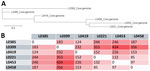
Figure 2. Core-genome phylogenetic relationships and single-nucleotide polymorphism (SNP) distance matrix from study of Trichosporon austroamericanuminfections among hospitalized patients, France, 2022–2024. A) Phylogenetic tree based on the core genome of...
The core genome consisting of 11,957 genes enabled construction of a phylogenetic tree from the extracted SNPs, which formed sequences with 542 nt sites and 196 parsimony-informative positions (Figure 2, panel A). Of the 542 sites, we observed 129 distinct site patterns, indicating a high level of sequence variation across the strains. Strains L0453 and L0221, separated by only 85 SNPs (Figure 2, panel B), were the closest in the tree. L0453, from the ICU air duct, also appeared near L0458, with a difference of 97 SNPs. In addition, we identified 7,668 shell genes, representing genes shared by a subset of strains, indicating genetic diversity beyond the core genome.
Whole-Genome SNP Analysis
We performed whole-genome SNP calling by mapping the Illumina reads of our strains against T. austroamericanum reference genome CBS 17435 (11). That method enabled us to include strain L0445, for which high-quality mapping data was available. Read mapping demonstrated high genome coverage quality across all 8 chromosomes and strict coverage exceeding 97% for all strains (minimum coverage 97.30% +SD 0.00368%) and reaching up to 99.98% (+SD 0.00043%).
Figure 3
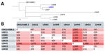
Figure 3. Whole-genome single-nucleotide polymorphism (SNP)–based phylogeny and distance matrix from study of Trichosporon austroamericanuminfections among hospitalized patients, France, 2022–2024. A) Whole-genome phylogenetic tree of the analyzed strains. Blue font...
The resulting whole-genome SNP distance matrix revealed patterns consistent with the core genome analysis (Figure 3, panel A). Distances between the CBS 17435 reference and the clinical strains ranged from 447 to 1,085 SNPs. The smallest pairwise distance was 105 SNPs between L0385 and L0399, which were 2 isolates collected within 3 months of each other from the same hospital, supporting their close genetic relationship. The second closest group consisted of L0221, L0453, and L0458, which clustered together despite being collected in different years (Table 1). Most other pairwise distances ranged from ≈400 to >1,100 SNPs, including between L0445 and the rest of the isolates (≈1,070–1,186 SNPs), suggesting that L0445 is genetically more distant, consistent with its location at a hospital site geographically distant from the others.
Mitochondrial Genome
As a complementary approach, we analyzed the mitochondrial genome to assess its potential for discriminating between strains. We used mapping to obtain mitochondrial genomes of ≈44 kb from 3 T. austroamericanum reference strains from the CNRMA and the L0445 strain, then generated genomes of other 6 genomes through de novo assembly (Appendix Figure 2). Of note, synteny was identical between the de novo assembled mitogenomes.
Figure 4
![Single-nucleotide polymorphism (SNP) map and phylogenetic analysis of mitogenomes from investigation of Trichosporon austroamericanum infections among hospitalized patients, France, 2022–2024. A) SNP map of mitochondrial genomes from T. austroamericanum isolates. The SNP alignment highlights 3 key positions in the mitochondrial genome (at positions 22240 in the cox3 gene, 31837 in the apocytochrome b gene, and 39897 in the trnL [tRNA-Leu] gene). B) Phylogenetic placement of clinical isolates (red font), the environmental strain (blue font), and strains from the National Reference Centre for Invasive Mycoses and Antifungals at Institut Pasteur (https://www.pasteur.fr). Scale bar indicates nucleotide substitutions per site.](/eid/images/25-0503-F4-tn.jpg)
Figure 4. Single-nucleotide polymorphism (SNP) map and phylogenetic analysis of mitogenomes from investigation of Trichosporon austroamericanum infections among hospitalized patients, France, 2022–2024. A) SNP map of mitochondrial genomes from ...
The alignment of those mitochondrial genomes against the de novo assembled L0221 sequence revealed 4 distinct mutational profiles, including 3 among Marseille strains (Figure 4). Those profiles included 3 key mutated positions in the mitochondrial genome of the cox3 (at position 22240), apocytochrome b (at position 31837), and trnL (tRNA-Leu) (at position 39897) genes. The mutations provide clear genetic distinctions among the strains. For instance, strain CNRMA15 exhibited a unique mutation, C at position 22240, whereas other strains, such as L0221, have a G at that position (Figure 4, panel A). That analysis underscores the value of mitochondrial genome sequencing for identifying and distinguishing between T. austroamericanum strains involved in this outbreak and future outbreaks.
The phylogenetic placement of mitochondrial genomes showed 4 distinct clades. L0221 and CNRMA15 formed a separate branch because of their unique SNP profiles. L0458 and L0453 were closely related, as seen in the core-genome analysis. Finally, L0385, L0419, L0399, L0445, CNRMA22, and CNRMA16 cluster together, and CNRMA 15 branches off the others (Figure 4, panel B).
With ≈20 pathogenic species, Trichosporon spp. are the second leading cause of Basidiomycota infections in humans (2). Although rare, Trichosporon infections in solid organ transplant patients have previously been reported and should be considered in cases of breakthrough infection or echinocandin therapeutic failure (20). In this case series, 67% of patients with invasive T. austroamericanum infection were kidney transplant recipients. For all kidney transplant recipients, the starting point was infection at the graft scar that occurred within 2–4 months of transplantation, suggesting a common source of contamination at the time of surgery. Of note, 2 patients in 2 different hospitals also had invasive T. austroamericanum infections, thus challenging the common source assumption. However, we did not observe any major changes in the antifungal susceptibility profiles that would have contradicted the idea of a common origin or that would have reflected an evolution of the strain over time (Table 2).
Figure 5
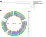
Figure 5. Maximum-likelihood phylogenetic tree and BLAST ring of isolates from a study of Trichosporon austroamericanuminfections among hospitalized patients, France, 2022–2024. A) Maximum-likelihood phylogenetic tree of the mitochondrial genome, showing...
T. austroamericanum was initially described in May 2024 by E.C. Francisco et al. (3). The first case we report appeared in July 2022, but because our database did not yet contain T. austroamericanum reference strains, the Trichosporon species were initially identified by MALDI-TOF mass spectrometry as T. inkin, a closely linked species. In the absence of a satisfactory reference genome, we focused on the T. inkin mitochondrial genome, the only genome available and well described in the literature (21). We observed considerable differences between the genomes of our strains, and the reference mitochondrial genome of T. inkin (Figure 5). The T. austroamericanum species described by Francisco et al. reinforced our analyses.
IGS1 is the region of interest for discriminating between different Trichosporon species (3,22,23). Comparing the rRNA IGS1 region nucleotide sequences between our strains with the CNRMA T. austroamericanum strains enabled us to confirm the result of mitochondrial genome analysis and revealed that our hospital was facing an emergence of T. austroamericanum. Moreover, the mitochondrial genome showed 3 different profiles within the T. austroamericanum strains isolated in Marseille and 3 key mutation positions. The appearance of spontaneous mutations has been described in the mitochondrial genome of certain phytopathogenic species, probably resulting from replication errors and the presence of mobile genetic elements (24).
We investigated a potential common origin through genomic analysis, as previously described (25). We produced complete genomes of T. austroamericanum by de novo WGS. Drawing inspiration from other epidemic investigations (26,27), we chose to target conserved sequences for a core-genome SNP typing approach (Figure 2). After publication of the T. austroamericanum CBS 17435 reference genome (11), we conducted a WGS and SNP analysis. Whole-genome SNP distances between the CBS 17435 reference strain from Brazil and our strains ranged from 447 to 1,186 SNPs. Of note, that range of differences was of the same order of magnitude as what we observed among the individual strains in our study, supporting the hypothesis of a polyclonal outbreak. Such an approach had already been used to identify clusters of 2 other basidiomycetes, T. asahii and Rhodotorula spp. (28,29).
No consensus regarding SNPs among fungal species exists because many strains are needed to study mutation rates, which is not always possible for rarer species. Thus, the cutoff point to determine whether strains are related seems to be set at <15 SNPs for Candida auris and 1,200 SNPs for other species, such as Rhodotorula mucilaginosa (28,30). Nevertheless, some strains in our study were genetically much closer. L0385 and L0399 differed by only 105 SNPs and were isolated from the same site within a short interval, suggesting a recent common origin or persistence from an environmental reservoir (Figure 3).
Phylogenetic analysis of the mitochondrial genome, core genome, and whole genome showed similar results, highlighting the benefits of those phylogenetic analyses in epidemiologic fungal investigations. Although the core-genome analysis was based on only a limited number (11,957) of genes, it enabled distinguishing the species and obtaining a phylogenetic placement like that from whole-genome analysis.
The hospital’s infection control team also conducted an environmental survey to identify a source of contamination, but investigations in the kidney transplant and nephrology departments did not reveal any common source of contamination. Investigations elsewhere have isolated Trichosporon species from the hospital environment. One published study identified 53 patients with positive T. asahii cultures in a hospital in Jamaica, 4 of whom were hospitalized in an ICU and had invasive T. asahii infection (5). After an environmental investigation in that hospital, 10 surface swab samples from the patient rooms showed T. asahii, including samples from drawers, bed rails, faucets, and sinks (5). In our investigation, T. austroamericanum was isolated (no. L0453) from an air sample from a pediatric ICU at hospital B, but no infection or colonization with that microorganism was noted among patients from that unit. We found no epidemiologic link between that unit and the reported clinical case-patients, all of whom were treated in other hospitals. The lack of an epidemiologic link suggests that T. austroamericanum was part of the hospital environment. In our study, the whole-genome SNP analyses showed 123–1,088 SNPs difference between the clinical strains and the environmental strain. However, that finding does not exclude the possibility of multiple clonally unrelated strains circulating and acquired within hospital environmental reservoir.
The results of this study demonstrate that core-genome analysis can effectively differentiate T. austroamericanum strains, revealing a polymorphic species. Furthermore, the mitochondrial genome shows strong potential as an excellent marker for intraspecies differentiation. That approach is particularly valuable in the absence of available annotated genomes for T. austroamericanum and was supplemented and reinforced by the release of the reference genome CBS 17435 (11).
In conclusion, our findings suggest that certain previously deposited sequences identified as T. inkin should be re-evaluated for T. austroamericanum to account for the emergence of this newly described species. In addition, this study underscores the need for vigilance regarding T. austroamericanum infections, including the potential for nosocomial involvement.
Ms. Burel is a PhD student in genomics and bioinformatics at the University of Aix Marseille, France. Her research interests include intrahost viral evolution, in particular and more broadly, the genomic evolution of microbial genomes.
Acknowledgments
The dataset generated for this study is available in the National Center for Biotechnology Information BioProject (https://www.ncbi.nlm.nih.gov/bioproject) database under accession nos. PRJDB20459 for mapped genomes and PRJNA1244900 for assemblies.
This work was supported by the government of France under the Investments for the Future program managed by the National Agency for Research, Mediterranean-Infection 10-IAHU-03, and was also supported by Région Provence-Alpes-Côte d’Azur and funding from FEDER PRIMMI (Fonds Européens de Développement Régional-Plateformes de Recherche et d’Innovation Mutualisées Méditerranée Infection), FEDER PA no. 0000320 PRIMMI.
Author contributions: conceptualization by E.M., S.R., and E.B.; materials and analysis tools contributed by E.M., E.B., C.S., V.M., J.S., J.So., M.D.O., and R.C.; data analysis by E.M. and E.B.; primary writing by E.M. and E.B.; review and editing by E.M., E.B., C.S., V.M., J.So., R.C., M.D.O., and S.R.; supervision by E.M. All authors read and agreed to the published version of the manuscript.
References
- Miceli MH, Díaz JA, Lee SA. Emerging opportunistic yeast infections. Lancet Infect Dis. 2011;11:142–51. DOIPubMedGoogle Scholar
- Menu E, Filori Q, Dufour JC, Ranque S, L’Ollivier C. A repertoire of the less common clinical yeasts. J Fungi (Basel). 2023;9:1099. DOIPubMedGoogle Scholar
- Francisco EC, Desnos-Ollivier M, Dieleman C, Boekhout T, Santos DWCL, Medina-Pestana JO, et al. Unveiling Trichosporon austroamericanum sp. nov.: a novel emerging opportunistic basidiomycetous yeast species. Mycopathologia. 2024;189:43. DOIPubMedGoogle Scholar
- Sugita T, Nishikawa A, Ichikawa T, Ikeda R, Shinoda T. Isolation of Trichosporon asahii from environmental materials. Med Mycol. 2000;38:27–30. DOIPubMedGoogle Scholar
- Fanfair RN, Heslop O, Etienne K, Rainford L, Roy M, Gade L, et al. Trichosporon asahii among intensive care unit patients at a medical center in Jamaica. Infect Control Hosp Epidemiol. 2013;34:638–41. DOIPubMedGoogle Scholar
- Menu E, Landier J, Prudent E, Ranque S, L’Ollivier C. Evaluation of 11 DNA automated extraction protocols for the detection of the 5 mains Candida species from artificially spiked blood. J Fungi (Basel). 2021;7:228. DOIPubMedGoogle Scholar
- Bolger AM, Lohse M, Usadel B. Trimmomatic: a flexible trimmer for Illumina sequence data. Bioinformatics. 2014;30:2114–20. DOIPubMedGoogle Scholar
- Lee S, Nguyen LT, Hayes BJ, Ross EM. Prowler: a novel trimming algorithm for Oxford Nanopore sequence data. Bioinformatics. 2021;37:3936–7. DOIPubMedGoogle Scholar
- Wick RR, Judd LM, Gorrie CL, Holt KE. Unicycler: Resolving bacterial genome assemblies from short and long sequencing reads. PLoS Comput Biol. 2017;13:
e1005595 . DOIPubMedGoogle Scholar - Simão FA, Waterhouse RM, Ioannidis P, Kriventseva EV, Zdobnov EM. BUSCO: assessing genome assembly and annotation completeness with single-copy orthologs. Bioinformatics. 2015;31:3210–2. DOIPubMedGoogle Scholar
- Francisco EC, Desnos-Ollivier M, Gerrits van den Ende B, Hagen F. De novo genome assembly and comparative genome analysis of the novel human fungal pathogen Trichosporon austroamericanum type-strain CBS 17435. Mycopathologia. 2025;190:33. DOIPubMedGoogle Scholar
- Li H, Durbin R. Fast and accurate short read alignment with Burrows-Wheeler transform. Bioinformatics. 2009;25:1754–60. DOIPubMedGoogle Scholar
- Grubaugh ND, Gangavarapu K, Quick J, Matteson NL, De Jesus JG, Main BJ, et al. An amplicon-based sequencing framework for accurately measuring intrahost virus diversity using PrimalSeq and iVar. Genome Biol. 2019;20:8. DOIPubMedGoogle Scholar
- Katoh K, Standley DM. MAFFT multiple sequence alignment software version 7: improvements in performance and usability. Mol Biol Evol. 2013;30:772–80. DOIPubMedGoogle Scholar
- Page AJ, Taylor B, Delaney AJ, Soares J, Seemann T, Keane JA, et al. SNP-sites: rapid efficient extraction of SNPs from multi-FASTA alignments. Microb Genom. 2016;2:
e000056 . DOIPubMedGoogle Scholar - Darling ACE, Mau B, Blattner FR, Perna NT. Mauve: multiple alignment of conserved genomic sequence with rearrangements. Genome Res. 2004;14:1394–403. DOIPubMedGoogle Scholar
- Minh BQ, Schmidt HA, Chernomor O, Schrempf D, Woodhams MD, von Haeseler A, et al. IQ-TREE 2: new models and efficient methods for phylogenetic inference in the genomic era. Mol Biol Evol. 2020;37:1530–4. DOIPubMedGoogle Scholar
- Cantarel BL, Korf I, Robb SMC, Parra G, Ross E, Moore B, et al. MAKER: an easy-to-use annotation pipeline designed for emerging model organism genomes. Genome Res. 2008;18:188–96. DOIPubMedGoogle Scholar
- Kalyaanamoorthy S, Minh BQ, Wong TKF, von Haeseler A, Jermiin LS. ModelFinder: fast model selection for accurate phylogenetic estimates. Nat Methods. 2017;14:587–9. DOIPubMedGoogle Scholar
- Almeida Júnior JN, Song ATW, Campos SV, Strabelli TMV, Del Negro GM, Figueiredo DSY, et al. Invasive Trichosporon infection in solid organ transplant patients: a report of two cases identified using IGS1 ribosomal DNA sequencing and a review of the literature. Transpl Infect Dis. 2014;16:135–40. DOIPubMedGoogle Scholar
- Liu Q, Wang X. Characterization and phylogenetic analysis of the complete mitochondrial genome of pathogen Trichosporon inkin (Trichosporonales: Trichosporonaceae). Mitochondrial DNA B Resour. 2021;6:803–5. DOIPubMedGoogle Scholar
- Araujo Ribeiro M, Alastruey-Izquierdo A, Gomez-Lopez A, Rodriguez-Tudela JL, Cuenca-Estrella M. Molecular identification and susceptibility testing of Trichosporon isolates from a Brazilian hospital. Rev Iberoam Micol. 2008;25:221–5. DOIPubMedGoogle Scholar
- Sugita T, Nakajima M, Ikeda R, Matsushima T, Shinoda T. Sequence analysis of the ribosomal DNA intergenic spacer 1 regions of Trichosporon species. J Clin Microbiol. 2002;40:1826–30. DOIPubMedGoogle Scholar
- Mendoza H, Perlin MH, Schirawski J. Mitochondrial inheritance in phytopathogenic fungi–everything is known, or is it? Int J Mol Sci. 2020;21:3883. DOIPubMedGoogle Scholar
- Douglas AP, Stewart AG, Halliday CL, Chen SCA. Outbreaks of fungal infections in hospitals: epidemiology, detection, and management. J Fungi (Basel). 2023;9:1059. DOIPubMedGoogle Scholar
- Aggelen HV, Kolde R, Chamarthi H, Loving J, Fan Y, Fallon JT III, et al. A core genome approach that enables prospective and dynamic monitoring of infectious outbreaks. Sci Rep. 2019;9:7808. DOIPubMedGoogle Scholar
- Menu E, Criscuolo A, Desnos-Ollivier M, Cassagne C, D’Incan E, Furst S, et al. Saprochaete clavata outbreak infecting cancer center through dishwasher. Emerg Infect Dis. 2020;26:2031–8. DOIPubMedGoogle Scholar
- Huang JJ, Chen XF, Tsui CKM, Pang CJ, Hu ZD, Shi Y, et al. Persistence of an epidemic cluster of Rhodotorula mucilaginosa in multiple geographic regions in China and the emergence of a 5-flucytosine resistant clone. Emerg Microbes Infect. 2022;11:1079–89. DOIPubMedGoogle Scholar
- Desnos-Ollivier M, Maufrais C, Pihet M, Aznar C, Dromer F; French Mycoses Study Group. Epidemiological investigation for grouped cases of Trichosporon asahii using whole genome and IGS1 sequencing. Mycoses. 2020;63:942–51. DOIPubMedGoogle Scholar
- Mitchell BI, Kling K, Bolon MK, Rathod SN, Malczynski M, Ruiz J, et al. Identifying Candida auris transmission in a hospital outbreak investigation using whole-genome sequencing and SNP phylogenetic analysis. J Clin Microbiol. 2024;62:
e0068024 . DOIPubMedGoogle Scholar
Figures
Tables
Cite This ArticleOriginal Publication Date: November 20, 2025
Table of Contents – Volume 31, Number 11—November 2025
| EID Search Options |
|---|
|
|
|
|
|
|
Please use the form below to submit correspondence to the authors or contact them at the following address:
Estelle Menu, Assistance Publique Hôpitaux de Marseille (AP-HM), Service de Parasitologie-Mycologie, 19-21 Blvd Jean Moulin, Marseille 13005, France
Top