Skip directly to site content Skip directly to page options Skip directly to A-Z link Skip directly to A-Z link Skip directly to A-Z link

Disclaimer: Early release articles are not considered as final versions. Any changes will be reflected in the online version in the month the article is officially released.

Volume 31, Number 11—November 2025

Research Letter

Fatal Tick-Borne Encephalitis in Unvaccinated Traveler from the United States to Switzerland, 2022

Chiara Scotti1, Gilbert Greub1Comments to Author , Yannis Ahmad, Simon Burgermeister, Giovanni Di Liberto, Ekkehard Hewer, Paola Vassallo, and Olivier Pantet1
Author affiliation: Lausanne University Hospital and University of Lausanne, Lausanne, Switzerland (C. Scotti, G. Greub, Y. Ahmad, S. Burgermeister, G. Di Liberto, E. Hewer, P. Vassallo, O. Pantet); Queen Square Institute of Neurology, London, UK (P. Vassallo); Leiden University Medical Centre, Leiden, the Netherlands (P. Vassallo); Stichting Epilepsie Instellingen Nederland, Heemstede, the Netherlands (P. Vassallo)

Main Article

Figure

Neuropathologic autopsy findings from cerebellum of unvaccinated traveler from the United States who died of tickborne encephalitis, Switzerland, 2022. A) shows Extensive lymphocytic infiltrate involving both leptomeninges and cerebellar parenchyma with depletion of the Purkinje cell layer (arrows). Scale bar represents 300 μm. B) The infiltrate consisted predominantly of cluster of differentiation 3 + T cell lymphocytes. Scale bar represents 300 μm. C) Low magnification illustrates the diffuse and extensive nature of the infiltrate also involving the white matter with perivascular accentuation (black arrows) and the dentate nucleus (white arrows). Scale bar represents 3 mm. Hematoxylin and eosin staining.

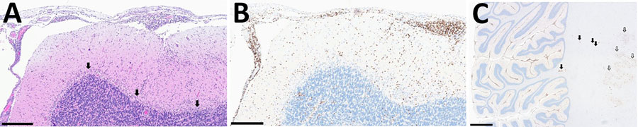
Figure. Neuropathologic autopsy findings from cerebellum of unvaccinated traveler from the United States who died of tickborne encephalitis, Switzerland, 2022. A) shows Extensive lymphocytic infiltrate involving both leptomeninges and cerebellar parenchyma with depletion of the Purkinje cell layer (arrows). Scale bar represents 300 μm. B) The infiltrate consisted predominantly of cluster of differentiation 3 + T cell lymphocytes. Scale bar represents 300 μm. C) Low magnification illustrates the diffuse and extensive nature of the infiltrate also involving the white matter with perivascular accentuation (black arrows) and the dentate nucleus (white arrows). Scale bar represents 3 mm. Hematoxylin and eosin staining.

Main Article

1These authors contributed equally to this article.

Page created: September 30, 2025
Page updated: November 20, 2025
Page reviewed: November 20, 2025
The conclusions, findings, and opinions expressed by authors contributing to this journal do not necessarily reflect the official position of the U.S. Department of Health and Human Services, the Public Health Service, the Centers for Disease Control and Prevention, or the authors' affiliated institutions. Use of trade names is for identification only and does not imply endorsement by any of the groups named above.
file_external