Case of Congenital Tularemia with Neuroinvasive Disease, Utah, USA
Brent D. Nelson

, Amara Finch, Krow Ampofo, Elizabeth L. Ryals, Andrew T. Pavia, Anne J. Blaschke, Jody L. Lin, Benjamin Kalm, Angie White, Kacy D. Nowak, Julian A. Villalba, Julu Bhatnagar, Bert Lopansri, and Elizabeth D. Knackstedt
Author affiliation: University of Utah School of Medicine, Salt Lake City, Utah, USA (B.D. Nelson, A. Finch, K. Ampofo, A.T. Pavia, A.J. Blaschke, J.L. Lin, B. Kalm, B. Lopansri, E.D. Knackstedt); Primary Children’s Hospital, Salt Lake City (E.L. Ryals); Bear River Health Department, Logan, Utah, USA (A. White); Utah Department of Health and Human Services, Salt Lake City (K.D. Nowak); Centers for Disease Control and Prevention, Atlanta, Georgia, USA (J.A. Villalba, J. Bhatnagar); Intermountain Medical Center, Murray, Utah, USA (B. Lopansri)
Main Article
Figure 1
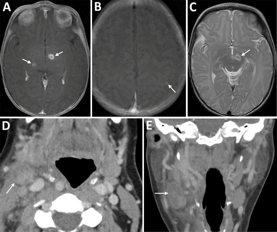
Figure 1. Imaging from infant and mother in case of congenital tularemia with neuroinvasive disease, Utah, USA. A, B) Axial T1 post-contrast images showing the infant’s initial magnetic resonance imaging findings of rim enhancing lesions near the left subthalamic nucleus and right inferior thalamus (arrows, panel A), as well as a punctate enhancing lesion in the left parietal lobe (arrow, panel B). C) Axial T2 image demonstrating T2 hyperintense edema along the margins of the largest lesion near the left subthalamic nucleus (arrow). D, E) Axial (D) and coronal (E) images from the mother’s computed tomography scan with intravenous contrast showing an enlarged, heterogeneous right cervical chain lymph node with inflammatory stranding in the adjacent soft tissues (arrows).
Main Article
Page created: November 18, 2025
Page updated: January 01, 2026
Page reviewed: January 01, 2026
The conclusions, findings, and opinions expressed by authors contributing to this journal do not necessarily reflect the official position of the U.S. Department of Health and Human Services, the Public Health Service, the Centers for Disease Control and Prevention, or the authors' affiliated institutions. Use of trade names is for identification only and does not imply endorsement by any of the groups named above.