Skip directly to site content Skip directly to page options Skip directly to A-Z link Skip directly to A-Z link Skip directly to A-Z link

Disclaimer: Early release articles are not considered as final versions. Any changes will be reflected in the online version in the month the article is officially released.

Volume 31, Number 2—February 2025
Synopsis

Two Human Cases of Fatal Meningoencephalitis Associated with Potosi and Lone Star Virus Infections, United States, 2020–2023

Charles Y. ChiuComments to Author , Raja Rama Godasi, Holly R. Hughes, Venice Servellita, Kafaya Foresythe, Asritha Tubati, Kelsey Zorn, Sukhman Sidhu, Michael R. Wilson, Sai Varun Bethina, Daniel Abenroth, Yee Cheng, Raymond Grams, Camilla Reese, Carlos Isada, and Neeharika Thottempudi
Author affiliation: Chan–Zuckerberg Biohub, San Francisco, California, USA (C.Y. Chiu); Abbott Pandemic Defense Coalition, Abbott Park, Illinois, USA (C.Y. Chiu, V. Servellita, K. Foresythe); University of California, San Francisco (C.Y. Chiu, V. Servellita, K. Foresythe, A. Tubati, K. Zorn, S. Sidhu, M.R. Wilson); St. Luke’s Boise Medical Center, Boise, Idaho, USA (R. Rama Godasi, D. Abenroth, Y. Cheng, R. Grams, C. Reese); Centers for Disease Control and Prevention, Fort Collins, Colorado, USA (H.R. Hughes); Wake Forest Baptist Medical Center, Winston-Salem, North Carolina, USA (S. Varun Bethina); Cleveland Clinic, Cleveland, Ohio, USA (C. Isada); University of Nevada, Reno, Nevada (N. Thottempudi)

Main Article

Figure

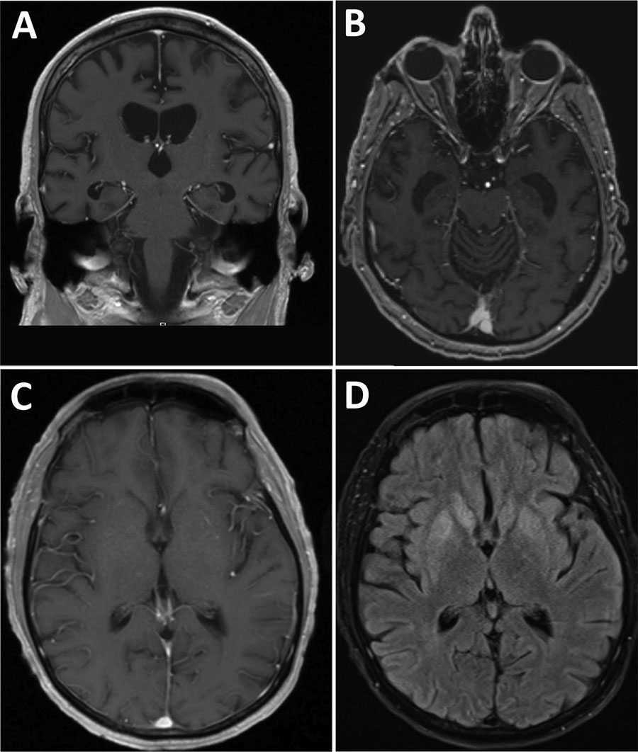
Brain magnetic resonance imaging scans from 2 patients with bunyavirus-associated meningoencephalitis, United States, 2020–2023. A, B) Case-patient 1 brain T1 postcontrast images of coronal (A) and axial (B) sections showing moderately enlarged ventricles and cerebral atrophy. C, D) Case-patient 2 brain T1 postcontrast (C) and T2 postcontrast fluid attenuated inversion recovery (D) images demonstrating bilateral basal ganglia hyperintensities with no contrast enhancement.

Figure. Brain magnetic resonance imaging scans from 2 patients with bunyavirus-associated meningoencephalitis, United States, 2020–2023. A, B) Case-patient 1 brain T1 postcontrast images of coronal (A) and axial (B) sections showing moderately enlarged ventricles and cerebral atrophy. C, D) Case-patient 2 brain T1 postcontrast (C) and T2 postcontrast fluid attenuated inversion recovery (D) images demonstrating bilateral basal ganglia hyperintensities with no contrast enhancement.

Main Article

Page created: December 20, 2024
Page updated: January 22, 2025
Page reviewed: January 22, 2025
The conclusions, findings, and opinions expressed by authors contributing to this journal do not necessarily reflect the official position of the U.S. Department of Health and Human Services, the Public Health Service, the Centers for Disease Control and Prevention, or the authors' affiliated institutions. Use of trade names is for identification only and does not imply endorsement by any of the groups named above.
file_external