Volume 31, Number 2—February 2025
Dispatch
Bjerkandera adusta Fungi as Causative Agent of Invasive Chronic Rhinosinusitis
Yuhei Kurata, Yoshifumi Kimizuka , Takashi Yaguchi, Kanshu Ito, Tetsuya Yamamoto, Yusuke Serizawa, Akira Kamiya, Takaaki Hamamoto, Taishi Sakima, Tomomi Tanigaki, Hiromi Edo, Yu Hongo, Akira Watanabe, Kazushi Suzuki, Terushige Toyooka, and Akihiko Kawana
Figure 1
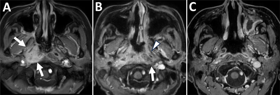
Figure 1. Chronological changes in lesions observed on contrast-enhanced fat-suppressed T1-weighted magnetic resonance imaging at the nasopharyngeal level in a patient in Japan with invasive chronic rhinosinusitis caused by Bjerkandera adusta fungi. A) Initial visit. Enhancement effects observed in the right nasopharynx, including the right torus tubarius and right prevertebral space (white arrows). B) Three months after the initial visit. Expansion of the enhancing lesion is seen, with enhancement extending to the left peritubal region (white arrowhead) and prevertebral space (white arrow). C) One year after the initial visit. The abnormal enhancement previously observed around both the torus tubarius and prevertebral space has regressed.
Page created: December 31, 2024
Page updated: January 31, 2025
Page reviewed: January 31, 2025
The conclusions, findings, and opinions expressed by authors contributing to this journal do not necessarily reflect the official position of the U.S. Department of Health and Human Services, the Public Health Service, the Centers for Disease Control and Prevention, or the authors' affiliated institutions. Use of trade names is for identification only and does not imply endorsement by any of the groups named above.