Volume 31, Number 3—March 2025
Research Letter
Urban Coatis (Nasua nasua) Exposure to Alphainfluenzavirus influenzae
Bruna Hermine de Campos, Jéssica de Souza Joaquim, Nadja Simbera Hemetrio, Lara Ribeiro de Almeida, Paula Cristina Senra Lima, Grazielle Cossenzo Florentino Galinari, Marcelo Coelho Lopes, Camila Issa Amaral, Gustavo Canesso Bicalho, Beatriz Senra Santos, Nágila Rocha Aguilar, Maria Isabel Maldonado Coelho Guedes, Danielle Ferreira de Magalhães Soares, Pedro Lúcio Lithg Pereira, Cíntia Aparecida de Jesus Pereira, Walter dos Santos Lima, Camila Stefanie Fonseca de Oliveira, Roselene Ecco, Erica Azevedo Costa, Zélia Inês Portela Lobato, and Marcelo Pires Nogueira de Carvalho
Figure
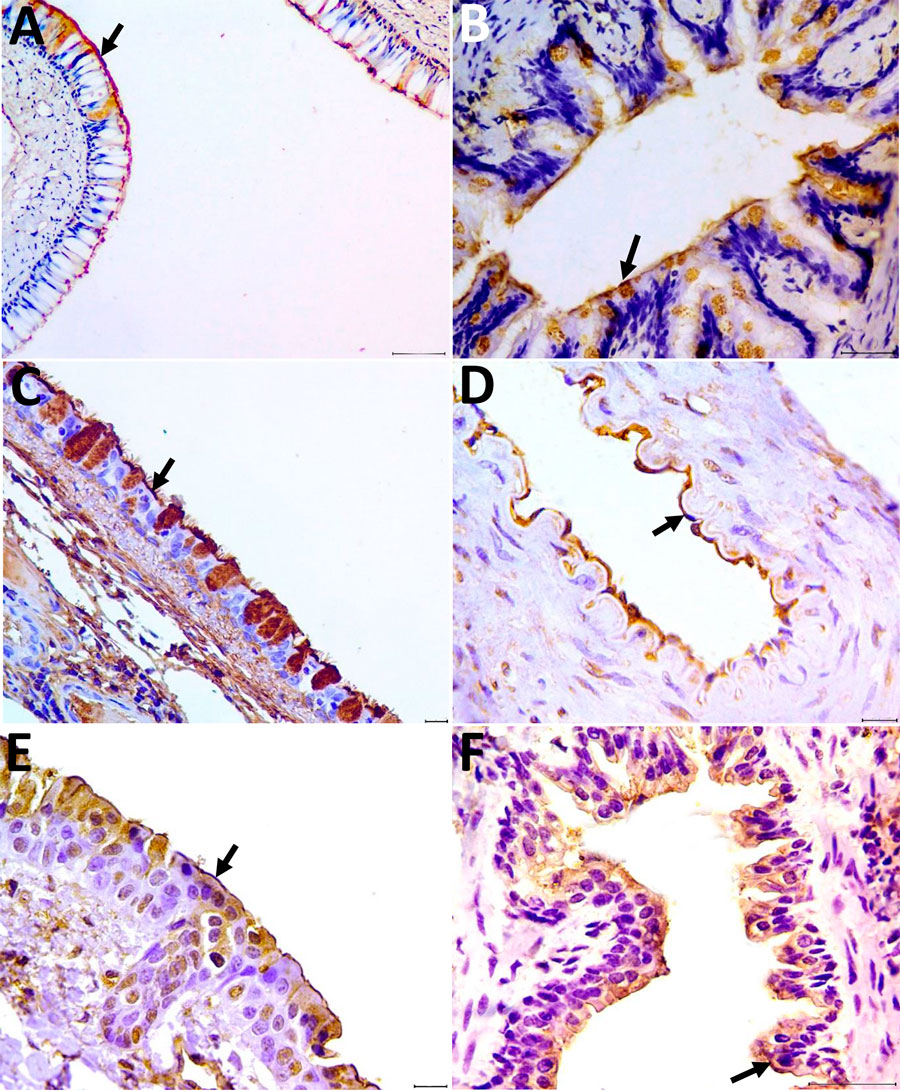
Figure. Detection of α-2,3 and α-2,6 receptors in tissues from the respiratory system of coatis (Nasua nasua), Brazil. A–C) Arrows indicate labeling of the α-2,3 receptor in the ciliated epithelium for the lectin Maackia amurensis II of the nasal concha (A), lung (bronchiole) tissue (B), and trachea (C). Scale bars = 100 µm in panel A, 50 µm in panel B, and 20 µm in panel C. D–F) Arrows indicate labeling of the α-2,6 receptor in the endothelium for Sambucus nigra lectin in the arteriole (D), rostral concha (E), and lung (bronchiole) (F). Scale bars = 20 µm in panels D and E, 50 µm in panel F. Tissue was counterstained with hematoxylin and revealed with diaminobenzidine chromogen.
Page created: February 04, 2025
Page updated: February 28, 2025
Page reviewed: February 28, 2025
The conclusions, findings, and opinions expressed by authors contributing to this journal do not necessarily reflect the official position of the U.S. Department of Health and Human Services, the Public Health Service, the Centers for Disease Control and Prevention, or the authors' affiliated institutions. Use of trade names is for identification only and does not imply endorsement by any of the groups named above.