Volume 31, Number 3—March 2025
Research
Effect of Prior Influenza A(H1N1)pdm09 Virus Infection on Pathogenesis and Transmission of Human Influenza A(H5N1) Clade 2.3.4.4b Virus in Ferret Model
Figure 2
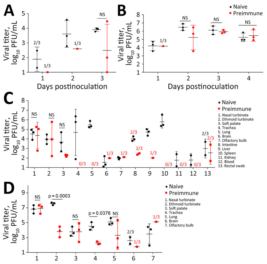
Figure 2. Virus shedding in study of the effect of prior influenza A(H1N1)pdm09 virus infection on pathogenesis and transmission of human influenza A(H5N1) clade 2.3.4.4b virus in ferret model. A, B) Nasal wash viral titers for influenza A(H5N1) Texas/37 virus (A) and influenza A(H7N9) Anhui/1 virus (B). C, D) Virus titers from tissues for Texas/37 H5N1 virus collected 3 days postinoculation (C) and Anhui/1 H7N9 virus collected 4 days postinoculation (D). Horizontal bars indicate median, dots indicate individual titers, whiskers indicate range of positive titers. Three naive and 3 pH1N1 preimmune ferrets were inoculated via respiratory inhalation with Texas/37 or Anhui/1 virus (Table). Nasal wash specimens (A, B) were collected daily. Virus titers were determined by standard plaque assay in MDCK cells. Tissue samples collected from nasal turbinate, ethmoid turbinate, soft palate, blood, and rectal swabs were reported in log10 PFU/mL. Tissues collected from lung, brain, olfactory bulb, intestines, liver, spleen, kidney were reported in log10 PFU/g. The limit of detection was 10 PFU per mL or g. Statistical analyses were performed using 2-way analysis of variance test when samples were positive for viral titers in all 3 inoculated animals; we considered p<0.05 statistically significant. When <3 inoculated ferrets had detectable virus, the detection frequency is indicated above the corresponding positions. Anhui/1, low pathogenicity influenza A(H7N9) A/Anhui/1/2013; NS, not statistically significant; Texas/37, highly pathogenic influenza A(H5N1) clade 2.3.4.4b A/Texas/37/2024.