Skip directly to site content Skip directly to page options Skip directly to A-Z link Skip directly to A-Z link Skip directly to A-Z link

Disclaimer: Early release articles are not considered as final versions. Any changes will be reflected in the online version in the month the article is officially released.

Volume 31, Number 3—March 2025
Dispatch

Donor-Derived Ehrlichiosis Caused by Ehrlichia chaffeensis from Living Donor Kidney Transplant

Michael J. Scolarici1Comments to Author , Daniel Kuehler1, Rebecca Osborn, Annie Doyle, Elizabeth K. Schiffman, Alex Garvin, Julian A. Villalba, Carmen J. Ramos, Christopher D. Paddock, Pallavi D. Annambhotla, Marissa Taylor, Johanna S. Salzer, Christopher Saddler, Carrie Thiessen, Raja Kandaswamy2, and Jon Odorico2
Author affiliation: University of Wisconsin–Madison School of Medicine and Public Health, Madison, Wisconsin, USA (M.J. Scolarici, C. Saddler, C. Thiessen, J. Odorico); University of Minnesota, Minneapolis, Minnesota, USA (D. Kuehler, A. Doyle, R. Kandaswamy); Wisconsin Department of Health Services, Madison (R. Osborn); Minnesota Department of Health, St. Paul, Minnesota, USA (E.K. Schiffman, A. Garvin); Centers for Disease Control and Prevention, Atlanta, Georgia, USA (J.A. Villalba, C.J. Ramos, C.D. Paddock, P.D. Annambhotla, M. Taylor, J.S. Salzer)

Main Article

Figure

Histopathologic and immunohistochemical staining of renal graft biopsy from donor-derived ehrlichiosis caused by Ehrlichia chaffeensis from living donor kidney transplant. We performed an immunohistochemical (IHC) assay using an immunoperoxidase technique, with naphthol fast-red substrate, light hematoxylin counterstaining, and an antibody raised against E. canis but known to cross-react with other Ehrlichia species, including E. chaffeensis and E. ewingii, among others. A) Interstitial peritubular mononuclear cell infiltrate (arrows). Hematoxylin and eosin (H&E) stain; original magnification ×400. B) Immunostaining of bacterial antigens from Ehrlichia species within 2 morulae in the cytoplasm of reactive endothelial cells of a renal venule (arrows). Original magnification ×630. C) Periglomerular focal interstitial mononuclear cell inflammation (arrows). H&E stain; original magnification ×400. D) Magnification of IHC assay of kidney biopsy indicating a granular immunostaining pattern within intracellular morulae (arrows). Original magnification ×1,000. E) Renal tubules displaying some features of acute tubular necrosis including epithelial cells with condensed chromatin and sloughing of cells into lumina. The tubular lumina are filled with sloughed off necrotic tubular epithelial cells (arrowheads). Some tubules show near complete luminal occlusion. Casts are not identified. H&E stain; original magnification ×630. F) Another intracellular morulae seen on kidney biopsy by IHC assay (arrow). Original magnification ×1,000.

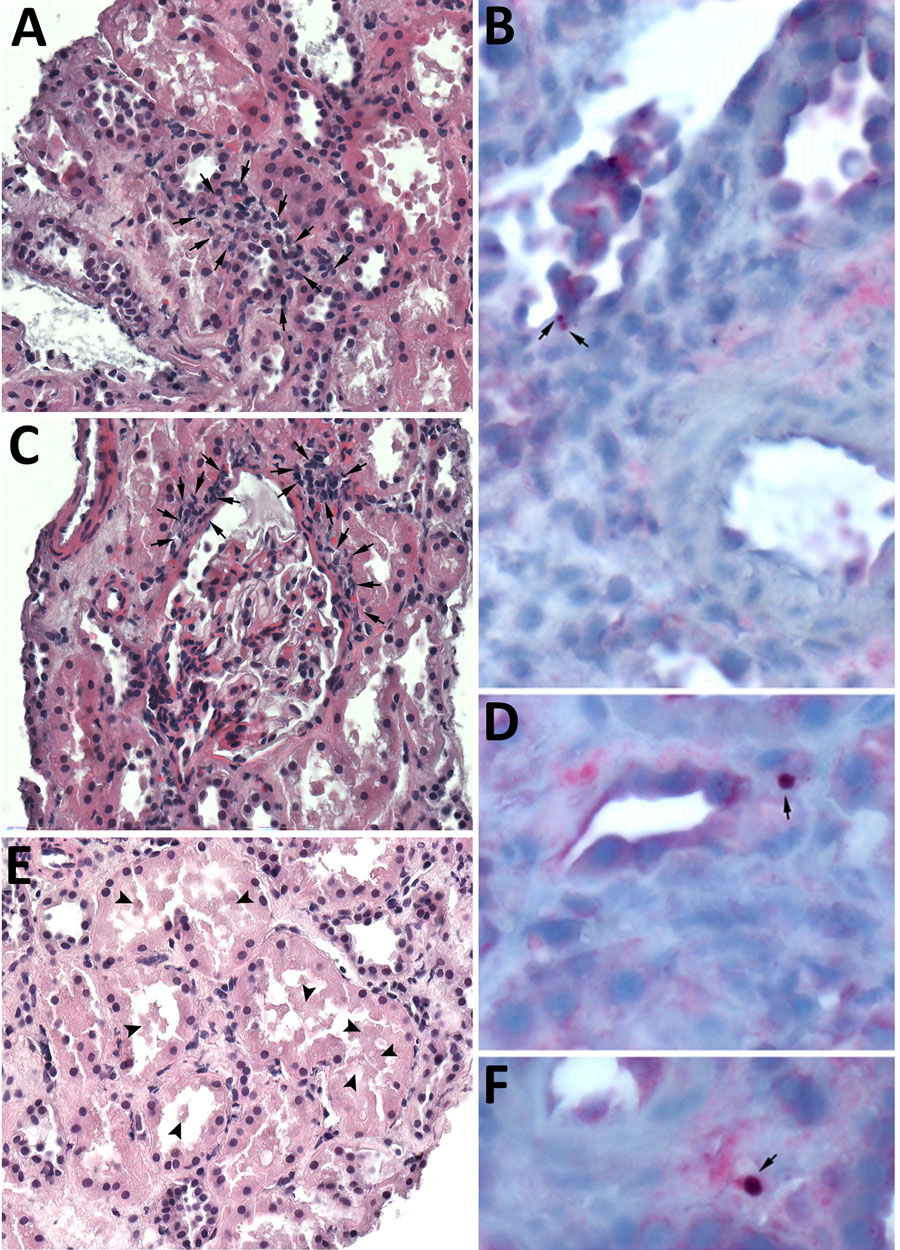
Figure. Histopathologic and immunohistochemical staining of renal graft biopsy from donor-derived ehrlichiosis caused by Ehrlichia chaffeensis from living donor kidney transplant. We performed an immunohistochemical (IHC) assay using an immunoperoxidase technique, with naphthol fast-red substrate, light hematoxylin counterstaining, and an antibody raised against E. canis but known to cross-react with other Ehrlichia species, including E. chaffeensis and E. ewingii, among others. A) Interstitial peritubular mononuclear cell infiltrate (arrows). Hematoxylin and eosin (H&E) stain; original magnification ×400. B) Immunostaining of bacterial antigens from Ehrlichia species within 2 morulae in the cytoplasm of reactive endothelial cells of a renal venule (arrows). Original magnification ×630. C) Periglomerular focal interstitial mononuclear cell inflammation (arrows). H&E stain; original magnification ×400. D) Magnification of IHC assay of kidney biopsy indicating a granular immunostaining pattern within intracellular morulae (arrows). Original magnification ×1,000. E) Renal tubules displaying some features of acute tubular necrosis including epithelial cells with condensed chromatin and sloughing of cells into lumina. The tubular lumina are filled with sloughed off necrotic tubular epithelial cells (arrowheads). Some tubules show near complete luminal occlusion. Casts are not identified. H&E stain; original magnification ×630. F) Another intracellular morulae seen on kidney biopsy by IHC assay (arrow). Original magnification ×1,000.

Main Article

1These first authors contributed equally to this article.

2These senior authors contributed equally to this article.

Page created: February 04, 2025
Page updated: February 24, 2025
Page reviewed: February 24, 2025
The conclusions, findings, and opinions expressed by authors contributing to this journal do not necessarily reflect the official position of the U.S. Department of Health and Human Services, the Public Health Service, the Centers for Disease Control and Prevention, or the authors' affiliated institutions. Use of trade names is for identification only and does not imply endorsement by any of the groups named above.
file_external