Skip directly to site content Skip directly to page options Skip directly to A-Z link Skip directly to A-Z link Skip directly to A-Z link

Disclaimer: Early release articles are not considered as final versions. Any changes will be reflected in the online version in the month the article is officially released.

Volume 31, Number 5—May 2025
Research Letter

Unexpected Zoonotic and Hybrid Schistosome Egg Excretion Patterns, Malawi, 2024

Angus M. O’Ferrall, Sekeleghe A. Kayuni, Lucas J. Cunningham, Peter Makaula, John Archer, Adam P. Roberts, Janelisa Musaya1, and J. Russell Stothard1Comments to Author 
Author affiliation: Liverpool School of Tropical Medicine, Liverpool, UK (A.M. O’Ferrall, S.A. Kayuni, L.J. Cunningham, J. Archer, A.P. Roberts, J.R. Stothard); Malawi-Liverpool-Wellcome Trust Clinical Research Programme, Queen Elizabeth Central Hospital, Blantyre, Malawi (S.A. Kayuni, P. Makaula, J. Musaya); School of Medicine and Oral Health, Kamuzu University of Health Sciences, Blantyre (S.A. Kayuni, J. Musaya); Wolfson Wellcome Biomedical Laboratories, Natural History Museum, London, UK (J. Archer)

Main Article

Figure

Morphologic Schistosoma spp. identification from 2 patient’s feces samples in an investigation of unexpected zoonotic and hybrid schistosome egg excretion patterns, Malawi, 2024. A) Typical S. haematobium (length 130 μm); B) typical S. mansoni (length 150 μm); C) atypical terminal-spined egg (length 188 μm); D) atypical terminal-spined egg (length 154 μm). Samples were stained with methylene blue glycerol solution using the Kato-Katz method (https://microbeonline.com/kato-katz-technique-principle-procedure-results). Atypical morphology (C,D) prompted closer molecular analysis for speciation, which revealed S. haematobium × S. mattheei hybrids.

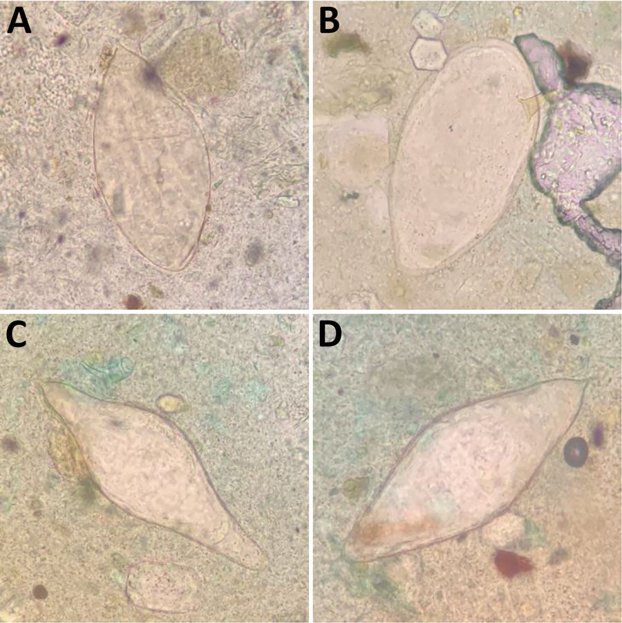
Figure. Morphologic Schistosoma spp. identification from 2 patient’s feces samples in an investigation of unexpected zoonotic and hybrid schistosome egg excretion patterns, Malawi, 2024. A) Typical S. haematobium (length 130 μm); B) typical S. mansoni (length 150 μm); C) atypical terminal-spined egg (length 188 μm); D) atypical terminal-spined egg (length 154 μm). Samples were stained with methylene blue glycerol solution using the Kato-Katz method (https://microbeonline.com/kato-katz-technique-principle-procedure-results). Atypical morphology (C,D) prompted closer molecular analysis for speciation, which revealed S. haematobium × S. mattheei hybrids.

Main Article

1These senior authors contributed equally to this article.

Page created: March 31, 2025
Page updated: April 17, 2025
Page reviewed: April 17, 2025
The conclusions, findings, and opinions expressed by authors contributing to this journal do not necessarily reflect the official position of the U.S. Department of Health and Human Services, the Public Health Service, the Centers for Disease Control and Prevention, or the authors' affiliated institutions. Use of trade names is for identification only and does not imply endorsement by any of the groups named above.
file_external