Volume 31, Number 9—September 2025
Etymologia
Apicoplast [ā′-pik-ō-plast]
Cite This Article
Citation for Media
The apicoplast is a unique organelle found in obligatory unicellular parasites called Apicomplexa due to a distinguished complex in their apex (top). The phylum Apicomplexa includes human pathogens, such as Plasmodium spp. that cause malaria and Toxoplasma spp. that cause toxoplasmosis, and prevalent veterinary parasites, such as Babesia and Eimeria spp.
The apicoplast was first identified in Toxoplasma parasites as a relict nonphotosynthetic chloroplast, a plastid, which is a term derived from the Greek plastos, meaning molded. The biological, evolutionary, and clinical consequences of that discovery were immediately apparent, and it was given the name apicoplast, a fusion of Apicomplexa and plastid. The name hints at the organelle’s unique evolutionary past. It was formed via secondary endosymbiosis, in which a unicellular protist engulfed another unicellular red alga and its chloroplast. Most Apicomplexan parasites retained that endosymbiont for metabolic purposes but lost all photosynthetic abilities. Few, like the genera of Cryptosporidium, lost the entire organelle. Of note, certain nonparasitic organisms related to Apicomplexa, like Chromera, still live as marine phototrophs, due to their photosynthetic plastid.
Figure
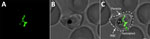
Figure. Visualization of the apicoplast organelle inside a malaria parasite. Microscopy image of a Plasmodium falciparumtransgenic parasite expressing a green fluorescent protein (GFP) fused to a transit peptide, which...
Regardless of photosynthesis, these plastids share similar metabolic pathways, have a small circular remnant genome, and are engulfed by no less than 4 distinct membranes (Figure). Perhaps more than anything, these membranes tell the evolutionary story of the apicoplast; much like a Russian Matryoshka doll, one organism is nested within another.
Acknowledgment
This work was supported by the Israel Science Foundation (ISF) under The Joint Canada-Israel Health Research Program (grant No. 3000/22 to A.F.). A.F. is supported by The Abisch-Frenkel Faculty Development Lectureship.
References
- Fichera ME, Roos DS. A plastid organelle as a drug target in apicomplexan parasites. Nature. 1997;390:407–9. DOIPubMedGoogle Scholar
- Janouškovec J, Horák A, Oborník M, Lukes J, Keeling PJ. A common red algal origin of the apicomplexan, dinoflagellate, and heterokont plastids. Proc Natl Acad Sci U S A. 2010;107:10949–54. DOIPubMedGoogle Scholar
- Köhler S, Delwiche CF, Denny PW, Tilney LG, Webster P, Wilson RJ, et al. A plastid of probable green algal origin in Apicomplexan parasites. Science. 1997;275:1485–9. DOIPubMedGoogle Scholar
- McFadden GI, Reith ME, Munholland J, Lang-Unnasch N. Plastid in human parasites. Nature. 1996;381:482. DOIPubMedGoogle Scholar
- Wilson RJ, Denny PW, Preiser PR, Rangachari K, Roberts K, Roy A, et al. Complete gene map of the plastid-like DNA of the malaria parasite Plasmodium falciparum. J Mol Biol. 1996;261:155–72. DOIPubMedGoogle Scholar
Figure
Cite This ArticleOriginal Publication Date: August 18, 2025
Related Links
Table of Contents – Volume 31, Number 9—September 2025
| EID Search Options |
|---|
|
|
|
|
|
|
Please use the form below to submit correspondence to the authors or contact them at the following address:
Anat Florentin, The Kuvin Center for the Study of Infectious and Tropical Diseases, Department of Microbiology and Molecular Genetics, Faculty of Medicine, The Hebrew University of Jerusalem, PO Box 12271, Jerusalem 91120, Israel
Top