Volume 31, Number 9—September 2025
Research Letter
Subarachnoid Neurocysticercosis Caused by Larval-Stage Taenia crassiceps Tapeworm, Slovenia
Barbara Šoba , Sandra Kolar, Albin Gačnik, Manca Radež, Timotej Petrijan, and Jana Rejc Marko
Figure 2
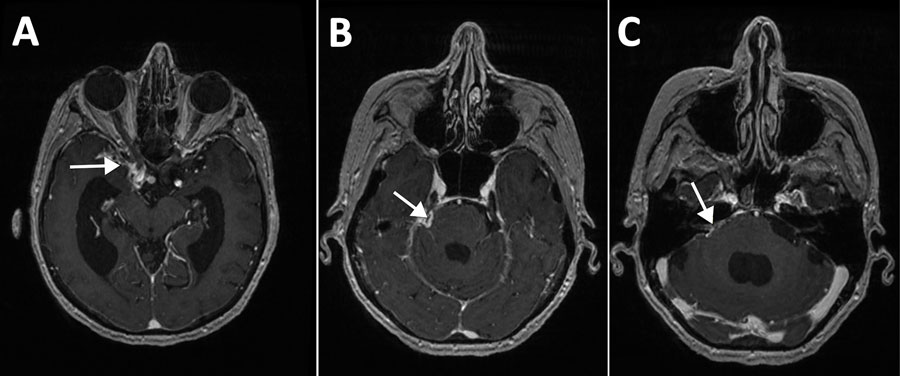
Figure 2. Magnetic resonance imaging of the brain in study of subarachnoid neurocysticercosis caused by larval-stage Taenia crassiceps tapeworm, Slovenia. Imaging shows pathological signal enhancement at the site of inflammation after contrast administration (white arrows) and a dilated ventricular system (hydrocephalus) as a result of impaired cerebrospinal fluid drainage in the right basal cisterns (A), right parapontine basal cistern (B), and right pontocerebellar angle (C).
Page created: July 11, 2025
Page updated: August 26, 2025
Page reviewed: August 26, 2025
The conclusions, findings, and opinions expressed by authors contributing to this journal do not necessarily reflect the official position of the U.S. Department of Health and Human Services, the Public Health Service, the Centers for Disease Control and Prevention, or the authors' affiliated institutions. Use of trade names is for identification only and does not imply endorsement by any of the groups named above.