Volume 32, Number 10—October 2026
Research Letter
Evaluation of Trichodysplasia Spinulosa Polyomavirus in Skin Biopsies, United States
Ivan M. Espinal, Gregory W. Charville, Lauren Lawrence, Ryanne A. Brown, Kerri E. Rieger, and Sebastian Fernandez-Pol
Figure
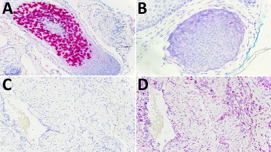
Figure. In situ hybridization (RNAscope) (5) for trichodysplasia spinulosa polyomavirus (TSPyV) in tissue samples, United States. A) Positive control tissue from a case of trichodysplasia spinulosa with staining for TSPyV. Original magnification ×200. B) peptidylprolyl isomerase B (PPIB) RNA probe staining on the same control tissue. Original magnification ×400. C) A representative case of inflammatory dermatosis with no staining for TSPyV RNA. Original magnification ×200. D) PPIB RNA probe staining on the same tissue in (C). Original magnification ×200.
References
- Lawrence L, Wang A, Charville G, Liu CL, Garofalo A, Alizadeh A, et al. Identification and confirmation via in situ hybridization of Merkel cell polyomavirus in rare cases of posttransplant cutaneous T-cell lymphoma. J Cutan Pathol. 2023;50:835–44. DOIPubMedGoogle Scholar
- Sadeghi M, Aaltonen LM, Hedman L, Chen T, Söderlund-Venermo M, Hedman K. Detection of TS polyomavirus DNA in tonsillar tissues of children and adults: evidence for site of viral latency. J Clin Virol. 2014;59:55–8. DOIPubMedGoogle Scholar
- van der Meijden E, Horváth B, Nijland M, de Vries K, Rácz EK, Diercks GF, et al. Primary polyomavirus infection, not reactivation, as the cause of trichodysplasia spinulosa in immunocompromised patients. J Infect Dis. 2017;215:1080–4.PubMedGoogle Scholar
- Borgogna C, Albertini S, Zavattaro E, Veronese F, Peruzzi L, van der Meijden E, et al. Primary trichodysplasia spinulosa polyomavirus infection in a kidney transplant child displaying virus-infected decoy cells in the urine. J Med Virol. 2019;91:1896–900. DOIPubMedGoogle Scholar
- Lawrence L, Wang A, Charville G, Toland A, Pinsky B, Natkunam Y, et al. Trichodysplasia spinulosa polyomavirus endothelial infection, California, USA. Emerg Infect Dis. 2022;28:1935–7. DOIPubMedGoogle Scholar
Page created: August 13, 2026
Page updated: September 28, 2026
Page reviewed: September 28, 2026
The conclusions, findings, and opinions expressed by authors contributing to this journal do not necessarily reflect the official position of the U.S. Department of Health and Human Services, the Public Health Service, the Centers for Disease Control and Prevention, or the authors' affiliated institutions. Use of trade names is for identification only and does not imply endorsement by any of the groups named above.