Skip directly to site content Skip directly to page options Skip directly to A-Z link Skip directly to A-Z link Skip directly to A-Z link

Disclaimer: Early release articles are not considered as final versions. Any changes will be reflected in the online version in the month the article is officially released.

Volume 32, Number 11—November 2026

Dispatch

Fatal Rickettsia parkeri Rickettsiosis in Immunocompromised Pediatric Patient, Georgia, USA, 2025

Stephanie Bellman1, Thomas G. Fox1, Sunil S. Raikar, Daniel John Bergsagel, Meredith Steuer, Kiran B. Hebbar, Jennifer Kasten-Metz, Sandor E. Karpathy, Christopher D. Paddock, and Kristy O. MurrayComments to Author 
Author affiliation: Children’s Healthcare of Atlanta, Atlanta, Georgia, USA (S. Bellman, T.G. Fox, S.S. Raikar, D.J. Bergsagel, M. Steuer, K.B. Hebbar, K.O. Murray); Emory University School of Medicine, Atlanta (S. Bellman, T.G. Fox, S.S. Raikar, D.J. Bergsagel, M. Steuer, K.B. Hebbar, K.O. Murray); Centers for Disease Control and Prevention, Atlanta (J. Kasten-Metz, S.E. Karpathy, C.D. Paddock)

Main Article

Figure 2

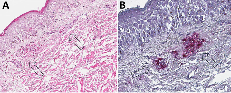
Immunohistochemistry and skin pathology from study of fatal Rickettsia parkeri rickettsiosis in immunocompromised pediatric patient, Georgia, USA, 2025. A) Punch biopsy specimen obtained from a rash lesion of the lower extremity of the patient demonstrating mild superficial dermal edema and small perivascular hemorrhages (arrows) without distinct inflammatory cell infiltrates. Hematoxylin and eosin stain; original magnification ×62.5. B) Immunohistochemical staining using an antibody reactive with spotted fever group Rickettsia spp., including Rickettsia parkeri, shows abundant staining of rickettsiae and rickettsial antigens in the lumens of the vessels, with invasion through the endothelium into the surrounding tissue (arrows). Immunoalkaline phosphatase with naphthol fast red and hematoxylin counterstain; original magnification ×62.5.

Figure 2. Immunohistochemistry and skin pathology from study of fatal Rickettsia parkeri rickettsiosis in immunocompromised pediatric patient, Georgia, USA, 2025. A) Punch biopsy specimen obtained from a rash lesion of the lower extremity of the patient demonstrating mild superficial dermal edema and small perivascular hemorrhages (arrows) without distinct inflammatory cell infiltrates. Hematoxylin and eosin stain; original magnification ×62.5. B) Immunohistochemical staining using an antibody reactive with spotted fever group Rickettsia spp., including Rickettsia parkeri, shows abundant staining of rickettsiae and rickettsial antigens in the lumens of the vessels, with invasion through the endothelium into the surrounding tissue (arrows). Immunoalkaline phosphatase with naphthol fast red and hematoxylin counterstain; original magnification ×62.5.

Main Article

1These first authors contributed equally to this article.

Page created: September 05, 2026
Page updated: October 16, 2026
Page reviewed: October 16, 2026
The conclusions, findings, and opinions expressed by authors contributing to this journal do not necessarily reflect the official position of the U.S. Department of Health and Human Services, the Public Health Service, the Centers for Disease Control and Prevention, or the authors' affiliated institutions. Use of trade names is for identification only and does not imply endorsement by any of the groups named above.
file_external