Disclaimer: Early release articles are not considered as final versions. Any changes will be reflected in the online version in the month the article is officially released.
Volume 32, Number 12—December 2026
Research
Identification of Zoonotic Pteropine orthoreovirus in Rousettus amplexicaudatus Fruit Bats, Indonesia
 , Diana Nurjanah, and Yustinus Maladan
Suggested citation for this article
Abstract
Pteropine orthoreovirus (PRV) is an emerging batborne virus with zoonotic potential increasingly linked to human respiratory illness. During 2020–2021, we sampled 182 fruit bats from animal markets and suppliers across Java, Indonesia, and identified species morphologically. We cultured rectal swab samples on Vero cells and sequenced samples showing cytopathic effects by using next-generation technology. We recovered 2 isolates from Rousettus amplexicaudatus bats sold for human consumption in Cianjur, West Java. Comparative analysis of 9 of 10 genomic segments showed the isolates were nearly identical (>99%–100%), except for the S2 segment (88.9%). Phylogenetic analysis indicated close clustering with Nelson Bay, Melaka, Kampar, Pulau, and Garut-69 bat-associated PRVs. We found PRV in R. amplexicaudatus bats in Indonesia, which highlights the zoonotic risk of wildlife trade. Continued surveillance of PRVs at the human–bat interface will be needed to assess the emergence of novel viruses and mitigate potential zoonotic threats.
Fruit bats, particularly large flying foxes (Pteropus vampyrus), are well documented as natural reservoirs of a wide range of viruses that have zoonotic potential, including herpesviruses, parvoviruses, polyomaviruses, paramyxoviruses, coronaviruses, and orthoreoviruses (1,2). Orthoreoviruses, specifically those classified as Pteropine orthoreovirus (PRV), have increasingly been associated with respiratory diseases in humans (3–5).
The zoonotic potential of batborne orthoreoviruses was first recognized in 2006, when a febrile illness along with respiratory symptoms were reported in a patient who had known exposure to a flying fox. The causative agent, later named Melaka virus, marked the first identification of a PRV responsible for human respiratory disease (6). After 2006, several genetically and antigenically related virus strains, such as Kampar, Sikamat, HK23629/07, and Miyazaki-Bali/2007, were isolated from patients who had similar respiratory tract infections (7–9). Those findings strongly support a role for PRVs as emerging zoonotic pathogens that have a capacity for interspecies transmission.
In 2010, a novel PRV strain, designated as Indonesia/2010, was isolated from a P. vampyrus bat imported from Indonesia to Italy (10). Whole-genome sequencing (WGS) revealed close genetic similarity to Melaka virus, reinforcing the hypothesis that Southeast Asia, particularly Indonesia, serves as a major ecologic reservoir for orthoreoviruses that have zoonotic potential. That discovery was followed by the identification of additional strains from Indonesia, such as Garut-69 (11), further highlighting the genetic diversity and ongoing circulation of PRVs in fruit bat populations.
A study in 2022 reported the isolation and characterization of a novel PRV strain from fruit bats in the Paguyaman region of Sulawesi, Indonesia; that strain was designated as Paguyaman orthoreovirus (12). In vitro and in vivo characterization of that virus revealed evidence of potential reassortment events with previously described Nelson Bay virus–related strains (8). Intranasal inoculation of laboratory mice with Paguyaman orthoreovirus resulted in severe pneumonia, underscoring its pathogenic potential in mammal hosts (12).
Mammal orthoreoviruses were first detected in Southeast Asia within fecal samples from P. vampyrus bats in Central Java Province, Indonesia (13). We report the detection of 2 PRV isolates in Rousettus amplexicaudatus bats collected in West Java, Indonesia. R. amplexicaudatus is a medium-sized fruit bat distributed throughout Indonesia. We conducted next-generation sequencing and phylogenetic analysis to determine associations between the 2 isolates and other orthoreoviruses.
Sample Collection
The Institutional Animal Care and Use Committee of the Indonesian Agency for Agricultural Research and Development approved all animal handling procedures (protocol nos. Balitbangtan/BB Litvet/M/01/2020 and Balitbangtan/BB Litvet/M/01/2021). We performed sampling nonlethally and obtained both rectal swab and blood serum samples from 182 bats. We placed rectal swab samples in virus transport medium consisting of Dulbecco’s modified Eagle medium (Thermo Fisher Scientific, https://www.thermofisher.com) supplemented with 10,000 U/mL penicillin/streptomycin, 250 μg/mL amphotericin B, and 10 mg/mL gentamycin (Thermo Fisher Scientific). We stored all samples at −20°C in a portable freezer and transported them to the Indonesia Research Center for Veterinary Science laboratory in Bogor, Indonesia.
Bat Species Identification
We identified bat species by using photographic records combined with established external morphologic assessments (14). Key morphometric parameters included forearm, tibia, hind limb, and ear lengths; overall body size and coloration; presence of a claw on the second wing digit; tail length; body mass; and distinctive features of the muzzle, tongue, and ear margins. We used those parameters to distinguish between bat species.
Virus Isolation, RNA Extraction, and WGS
We extracted virus RNA by using the QIAamp Viral RNA Mini Kit (QIAGEN, https://www.qiagen.com), according to the manufacturer’s protocol. We preserved the RNA extracts at −20°C for reverse transcription PCR (RT-PCR). We screened total RNA extracts from collected samples for influenza A virus (15) and for RNA-dependent RNA polymerase (RdRp) and spike genes of bat coronaviruses by RT-PCR (16). In addition, we screened for SARS-CoV-2 by using the SensiFAST Probe No-ROX One-Step Real-Time PCR Kit (Bioline, https://www.bioline.com) on an Applied Biosystems 7300 Real-Time PCR System (Thermo Fisher Scientific). The SARS-CoV-2 assay targeted the nucleocapsid (N) 1 and N2 gene regions, and we used the human RNase P gene as an internal control, according to the manufacturer’s protocol. We used the SARS-CoV-2 assay for qualitative real-time RT-PCR to detect the presence of the virus; the assay was not intended for virus load quantification.
After screening, we selected a subset of samples that tested positive for a virus and performed virus isolation through a blinded cell culture procedure. We inoculated Vero cell monolayers with each sample separately and incubated the cells at 37°C in a humidified 5% CO2 atmosphere under standard cell culture conditions. We monitored cultures daily for the appearance of cytopathic effects (CPEs). Among the cultured samples, we selected 2 isolates exhibiting prominent CPEs for further genomic analysis. We reextracted virus RNA directly from clarified culture supernatants and performed WGS using next-generation sequencing technology. A commercial sequencing service provider performed the library preparation and sequencing by using the Illumina NextSeq 550 platform with paired-end 150-bp reads. We used the resulting sequencing data for virus genome assembly and downstream molecular characterization. In addition, we also performed RT-PCR targeting the small (S) 1 and S2 genome segments as previously described (11) to confirm the presence of those segments in the isolates (Appendix Figure 1).
Bioinformatics Analysis
We detected and quantified animal virus pathogens from short-read sequencing data by using EsViritu version 3.1.0 (https://github.com/cmmr/EsViritu). EsViritu is a sensitive and specific bioinformatics pipeline designed to explore the presence, absence, and diversity of viruses within and between metagenomic or clinical samples. The database used by EsViritu includes all human and animal viruses available in GenBank. The output of the pipeline consists of consensus genomes or segments of detected viruses along with an interactive table that includes visualizations of read coverage profiles and accession numbers of identified viruses (M.J. Tisza et al., unpub. data, https://doi.org/10.64898/2026.09.29.755457).
We analyzed the extracted fasta data obtained in this study together with the genetic data available in the National Center for Biotechnology Information Virus database (https://www.ncbi.nlm.nih.gov/labs/virus/vssi) for orthoreoviruses. We performed multiple gene alignments and analysis of amino acids by using BioEdit version 7 (http://www.mbio.ncsu.edu/BioEdit). We generated phylogenetic trees by using the maximum likelihood method (1,000 bootstraps) and the Tamura-Nei algorithm in MEGA version 12 (http://www.megasoftware.net). We deposited all nucleotide sequence data generated in this study in GenBank (BankIt2989076, accession nos. PX113089–106).
Sample Collection and Bat Species Identification
Figure 1

Figure 1. Bat collection sites for study of zoonotic Pteropine orthoreovirus in Rousettus amplexicaudatusfruit bats, Indonesia. Map shows Central Java (Surakarta city and Magelang Regency), the Special Region...
We collected a total of 182 bats during 2 sampling periods (126 in 2020 and 56 in 2021) from animal markets and bat suppliers operating in several locations across Central Java (Surakarta City and Magelang Regency), Yogyakarta Special Region, and West Java (Bogor City and Cianjur Regency) (Figure 1). We taxonomically classified the collected bats into 4 species: Cynopterus brachyotis (n = 45), Macroglossus minimus (n = 5), R. amplexicaudatus (n = 96), and Pteropus vampyrus (n = 36).
Virus Isolation, WGS, and Bioinformatics Analysis
Figure 2
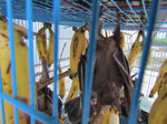
Figure 2. Rousettus amplexicaudatus bats, a frugivorous species collected during study of zoonotic Pteropine orthoreovirus in R. amplexicaudatus fruit bats, Indonesia.
Two virus isolates, designated PRV/SJD-K30/ID/2021 and PRV/SJD-K50/ID/2021, exhibited CPEs in Vero cell cultures. Therefore, we conducted further analyses of those isolates. Both samples were derived from fruit bats identified morphologically as R. amplexicaudatus (Figure 2). Those 2 bats were obtained from a bat collector operating in a residential area in Cianjur, West Java, Indonesia, where bats are sold for human consumption.
We screened samples for influenza A virus, bat coronavirus RdRp and spike genes, and SARS-CoV-2 N1 and N2 gene regions (Table 1). All assays were negative except for the bat coronavirus RdRp screening assay, which yielded positive results in both PRV/SJD-K30/ID/2021 and PRV/SJD-K50/ID/2021. We obtained PRV/SJD-K30/ID/2021 from a live male bat (live at the time of sampling) and PRV/SJD-K50/ID/2021 from a dead male bat; both isolates showed identical screening results (Table 2). Both isolates yielded positive results in the bat coronavirus RdRp screening assay and produced CPE in Vero cell cultures. However, next-generation sequencing did not confirm coronavirus genomic sequences in the samples. Instead, sequence analysis identified a novel PRV, indicating the initial RdRp screening by RT-PCR produced nonspecific amplification that was not corroborated by WGS.
Figure 3

Figure 3. Schematic representation of the complete orthoreovirus virion and 9 genomic segments sequenced in study of zoonotic Pteropine orthoreovirus in R. amplexicaudatusfruit bats, Indonesia. Both PRV/SJD-K30/ID/2021 and...
We reconstructed genomes of the isolated PRVs according to WGS results (Figure 3). PRV genomes comprise 10 double-stranded RNA segments that are classified into 3 large (L), 3 medium (M), and 4 S segments. Each segment encodes >1 protein essential for virus replication, structure, or host interaction. However, in our study, we only recovered 9 segments, indicating incomplete genome acquisition possibly caused by low RNA abundance, degradation, or technical limitations during amplification or sequencing. S1 was not recovered. We further assessed recovery by nested PCR targeting S1 and S2 gene segments by using RNA extracted from Vero cell culture isolates at passage numbers 0 and 1. Although the S2 segment was successfully amplified, the S1 segment yielded only a weak and barely detectable amplicon (Appendix Figure 1). The weak S1 amplification is consistent with low template abundance and might explain the inability to recover all genome segments during next-generation sequencing.
We reconstructed the orthoreovirus virions, which contained 9 genomic segments ranging in size from 1,076 to 3,843 bp (Figure 3). The absence of segment S1, commonly associated with the σC protein involved in cell attachment and host specificity, might have affected downstream phenotypic assessments but did not hinder classification of the isolates within the PRV group. Comparative alignment of segment sizes and gene arrangements further supports the identity of the isolated viruses as a member of the PRV species.
Figure 4

Figure 4. Maximum-likelihood phylogenetic tree of Pteropine orthoreovirus genomic L1 segment found in Rousettus amplexicaudatusfruit bats, Indonesia. Black circles indicate orthoreovirus isolates PRV/SJD-K30/ID/2021 and PRV/SJD-K50/ID/2021 identified in this...
Figure 5

Figure 5. Maximum-likelihood phylogenetic tree of Pteropine orthoreovirus genomic L2 segment found in Rousettus amplexicaudatusfruit bats, Indonesia. Black circles indicate orthoreovirus isolates PRV/SJD-K30/ID/2021 and PRV/SJD-K50/ID/2021 identified in this...
Figure 6

Figure 6. Maximum-likelihood phylogenetic tree of Pteropine orthoreovirus genomic L3 segment found in Rousettus amplexicaudatusfruit bats, Indonesia. Black circles indicate orthoreovirus isolates PRV/SJD-K30/ID/2021 and PRV/SJD-K50/ID/2021 identified in this...
Figure 7

Figure 7. Maximum-likelihood phylogenetic tree of Pteropine orthoreovirus genomic M1 segment found in Rousettus amplexicaudatusfruit bats, Indonesia. Black circles indicate orthoreovirus isolates PRV/SJD-K30/ID/2021 and PRV/SJD-K50/ID/2021 identified in this...
Figure 8

Figure 8. Maximum-likelihood phylogenetic tree of Pteropine orthoreovirus genomic M2 segment found in Rousettus amplexicaudatusfruit bats, Indonesia. Black circles indicate orthoreovirus isolates PRV/SJD-K30/ID/2021 and PRV/SJD-K50/ID/2021 identified in this...
Figure 9

Figure 9. Maximum-likelihood phylogenetic tree of Pteropine orthoreovirus genomic M3 segment found in Rousettus amplexicaudatusfruit bats, Indonesia. Black circles indicate orthoreovirus isolates PRV/SJD-K30/ID/2021 and PRV/SJD-K50/ID/2021 identified in this...
Figure 10

Figure 10. Maximum-likelihood phylogenetic tree of Pteropine orthoreovirus genomic S2 segment found in Rousettus amplexicaudatusfruit bats, Indonesia. Black circles indicate orthoreovirus isolates PRV/SJD-K30/ID/2021 and PRV/SJD-K50/ID/2021 identified in this...
Figure 11

Figure 11. Maximum-likelihood phylogenetic tree of Pteropine orthoreovirus genomic S3 segment found in Rousettus amplexicaudatusfruit bats, Indonesia. Black circles indicate orthoreovirus isolates PRV/SJD-K30/ID/2021 and PRV/SJD-K50/ID/2021 identified in this...
Figure 12

Figure 12. Maximum-likelihood phylogenetic tree of Pteropine orthoreovirus genomic S4 segment found in Rousettus amplexicaudatusfruit bats, Indonesia. Black circles indicate orthoreovirus isolates PRV/SJD-K30/ID/2021 and PRV/SJD-K50/ID/2021 identified in this...
We created phylogenetic trees for each of the 9 gene segments (Figures 4,5,6,7,8,9,10,11,12). Phylogenetic analyses of segments L1–L3, M1–M3, and S2–S4 showed that PRV/SJD-K30/ID/2021 and PRV/SJD-K50/ID/2021 consistently clustered with other bat-associated orthoreoviruses, including Nelson Bay (China), Cangyuan (China), Melaka (Malaysia), Kampar (Malaysia), and Pulau orthoreoviruses, as well as PRV (Malaysia) and PRV Garut-69 (Indonesia). Phylogenetic trees of L2, L3, M1, M3, S2, and S3 segments indicated that PRV/SJD-K30/ID/2021 and PRV/SJD-K50/ID/2021 were most closely related to the PRV Garut-69 strain isolated from Pteropus vampyrus bats, which was previously associated with human infections in Indonesia.
We generated heat maps to compare the 9 segments of PRV/SJD-K30/ID/2021 and PRV/SJD-K50/ID/2021 with sequences from other viruses (Appendix Figures 2–10). L1 segments exhibited 100% nucleotide identity with each other. Both isolates showed the highest similarity to PRVs, particularly Pulau (94.6%) and Melaka (94.1%) reoviruses, followed by Cangyuan orthoreovirus (91.7%). In contrast, identities with avian, reptile, and non-PRV mammal orthoreoviruses were much lower (≈27%–35%). The L1 sequences firmly place the 2 isolates within the PRV group. L2 gene segments, which encode capsid proteins, were 100% identical between the 2 isolates. Both isolates showed the highest identities with Pulau (93.3%) and Melaka (92.5%) reoviruses, followed by Cangyuan orthoreovirus (91.6%). Non-PRV reference sequences showed <65% identity. Thus, the L2 segment demonstrated a highly conserved backbone consistent with the PRV lineage. The L3 gene segments of the 2 isolates were 100% identical. The closest reference sequences were Melaka (78.9%), Pulau (78.7%), and Cangyuan (78.1%) orthoreoviruses, whereas other orthoreoviruses showed only 27%–50% identity. Those findings indicate the L3 segment of the 2 isolates clusters tightly within the PRV group.
M1 gene segments from PRV/SJD-K30/ID/2021 and PRV/SJD-K50/ID/2021 shared 99.1% identity. We detected the highest external similarities between PRV/SJD-K30/ID/2021 and Pulau (77.3%), Melaka (77.1%), and Cangyuan (77.0%) orthoreoviruses. PRV/SJD-K50/ID/2021 showed lower similarities with Pulau (76.8%), Melaka (76.5%), and Cangyuan (76.4%) orthoreoviruses. Those findings suggest the M1 segment has strong PRV affinity and shows minimal divergence between the 2 isolates from Indonesia.
The M2 gene segments, which encode outer capsid proteins, were 100% identical between the 2 isolates. The closest matches were Pulau (82.2%) and Melaka (82.0%) orthoreoviruses, whereas the match with Cangyuan orthoreovirus was slightly lower (69.8%). That pattern suggests indicates the conserved outer capsid determinants of the 2 isolates are similar to those of Pulau and Melaka viruses. In addition, the M3 segment of the 2 isolates shared 100% identity. The highest sequence similarities were with Pulau and Melaka orthoreoviruses (85.8% each), followed by Cangyuan orthoreovirus (85.0%). Thus, the M3 segment is strongly conserved within PRVs.
Unlike the other segments, the nucleotide identity of S2 gene segments between PRV/SJD-K30/ID/2021 and PRV/SJD-K50/ID/2021 decreased to 88.9%. Nevertheless, both isolate sequences remained closest to other PRVs, including Pulau (83.3%), Melaka (81.5%–81.8%), and Cangyuan (81.7%) orthoreoviruses. The decreased identity between the 2 isolates likely indicates an intra-PRV reassortment event in the S2 segment. However, the presence of numerous ambiguous nucleotide positions in 1 of the sequences might have influenced the estimated sequence identity and limited the strength of that inference.
The S3 gene segments of the 2 isolates were nearly identical (99.9%). The highest similarities were to Pulau (78.1%–78.2%), Melaka (77.0%), and Cangyuan (76.3%–76.4%) orthoreoviruses. No evidence of reassortment was observed in this segment.
The S4 gene segments of the 2 isolates were 100% identical. The highest identities were with Melaka orthoreovirus (86.7%), followed by Cangyuan (85.2%) and Pulau (83.3%) orthoreoviruses, suggesting that the S4 segment, which plays a role in viral entry, is highly conserved among PRVs.
Taken together, analyses of all 9 segments consistently place PRV/SJD-K30/ID/2021 and PRV/SJD-K50/ID/2021 within the PRV clade, and the closest affinities were to Melaka, Pulau, and Cangyuan orthoreoviruses. The 2 isolates from Indonesia were nearly identical across the genome (>99%–100%), except for the S2 segment, which declined to ≈89%. That pattern strongly suggests reassortment within locally circulating PRVs that specifically involves the S2 segment, which encodes an inner capsid structural protein. Our findings support the existence of active PRV circulation in Indonesia with >2 divergent S2-associated lineages embedded in an otherwise conserved PRV genomic background.
R. amplexicaudatus, a medium-sized frugivorous bat species belonging to the order Chiroptera, suborder Megachiroptera, and family Pteropodidae, is 1 of 4 recognized Rousettus species native to Indonesia. Locally known as codot or regular nyap bats, R. amplexicaudatus bats are widely distributed across the archipelago of Indonesia, particularly in Central Java, Yogyakarta, and West Java, including areas such as Cianjur (17). The bats commonly roost in a range of habitats, including natural caves, forested areas, abandoned buildings, and other anthropogenic structures, which reflects its high adaptability to both natural and semiurban environments.
Ecologically, R. amplexicaudatus bats play a critical role as pollinators and seed dispersers and contribute to ecosystem regeneration and biodiversity. However, the frequent proximity of the bats to human settlements, their presence in local wildlife markets, and human consumption as bushmeat has increased concern regarding zoonotic disease transmission. Recent surveillance studies have identified R. amplexicaudatus bats as a potential reservoir for various emerging viruses, such as bat-associated coronaviruses, hantavirus, and filovirus (18–20).
Despite wide distribution across Indonesia (12,21–23), R. amplexicaudatus bats have not been reported as hosts for orthoreoviruses (12,13). That absence of documentation might reflect a substantial knowledge gap in the country’s bat virome surveillance, which has historically focused on more widely recognized zoonotic agents, such as coronaviruses, filoviruses, and henipaviruses. Because R. amplexicaudatus bats are ecologically ubiquitous and frequently interface with humans through hunting, trade, and consumption, increased surveillance of orthoreovirus infection will be needed.
We identified 2 bat-associated orthoreovirus isolates, PRV/SJD-K30/ID/2021 and PRV/SJD-K50/ID/2021, in R. amplexicaudatus bats from Indonesia. Despite successful recovery and phylogenetic characterization of 9 of 10 genomic segments from those isolates, the S1 segment was not detected in either isolate. The S1 segment encodes the σC outer capsid protein, which is a viral attachment protein that forms homotrimers and plays critical roles in cell tropism and virus dissemination pathways in vivo. The absence of that segment in our assay is likely attributed to its high sequence variability (24), which poses challenges for primer annealing and accurate de novo sequence assembly, particularly when nontargeted metagenomic approaches are used. The S1 segment is known to be among the most genetically diverse segments in PRVs, often resulting in poor homology with reference sequences and inefficient read mapping during genome reconstruction. In addition, the potential low abundance or degradation of S1 RNA, especially within rectal swab sample–derived field samples, might have further limited its detection. Technical limitations related to cDNA synthesis, random priming efficiency, and sequencing bias might also have contributed to failed recovery of this segment. Those findings underscore the need for segment-specific enrichment strategies or targeted amplification protocols in future surveillance efforts to obtain complete orthoreovirus genome profiles, particularly for the highly variable S1 segment.
Phylogenetic analyses of all 9 genomic segments demonstrated consistent clustering within the PRV lineage, indicating a conserved genomic organization and a shared evolutionary origin. Those findings highlight the close genetic relationship and potential zoonotic relevance of the isolates. Furthermore, the geographic clustering of the 2 isolates with other PRVs from Southeast Asia and Oceania suggests regional virus dissemination, possibly promoted by bat migration or wildlife trade networks (25,26). Both isolates formed distinct clades and showed no phylogenetic association with avian, reptile, or mammal orthoreoviruses, thereby, reinforcing their classification within the batborne PRV group. Phylogenetic analyses also revealed that PRV/SJD-K30/ID/2021 and PRV/SJD-K50/ID/2021 consistently clustered alongside Nelson Bay (China) (8), Cangyuan (China) (27), Melaka (Malaysia) (4,28), Kampar (Malaysia) (5), and Pulau (29) orthoreoviruses, as well as PRV (Malaysia) and PRV Garut-69 (Indonesia). The 2 isolates are closely related to the Garut-69 strain from P. vampyrus, indicating a shared evolutionary lineage and potential zoonotic risk.
The segmented nature of the PRV genome promotes genetic reassortment, a mechanism by which viruses acquire novel combinations of gene segments (30). Those events are known drivers of virus adaptation and host range expansion. The observed genomic constellation of PRV/SJD-K30/ID/2021 and PRV/SJD-K50/ID/2021, which share identity with diverse regional isolates, raises the possibility of reassortment between endemic strains within bat populations that could lead to the emergence of viruses with altered pathogenic or zoonotic potential.
The phylogenetic proximity of PRV/SJD-K30/ID/2021 and PRV/SJD-K50/ID/2021 to Melaka virus, a strain associated with confirmed human respiratory infections (4,28), and to Garut-69, previously isolated in Indonesia, underscores the public health relevance of those findings. Because of the ongoing human–bat interface in wildlife markets and culinary settings across Indonesia (31,32), surveillance of batborne orthoreoviruses and their potential for interspecies transmission is imperative.
In conclusion, we found molecular evidence of PRV infection in R. amplexicaudatus bats in Indonesia, which expands the known host range of PRVs in Southeast Asia. The successful isolation and genomic characterization of 2 virus isolates (PRV/SJD-K30/ID/2021 and PRV/SJD-K50/ID/2021) from bats sold for human consumption in Cianjur, West Java, underscore the ongoing risk posed by unregulated wildlife trade and close human–bat interactions. Phylogenetic analysis revealed that both isolates are closely related to previously identified zoonotic strains, such as Melaka virus and Garut-69, suggesting a shared evolutionary origin and potential for interspecies transmission. The segmented nature of the PRV genome and the observed genetic constellation indicate historical reassortment events, further highlighting the dynamic evolution of orthoreoviruses within bat populations. Collectively, our findings reinforce Indonesia’s ecologic role as a hotspot for PRV diversity and highlight the urgent need for continuous, integrated genomic surveillance at the human–wildlife interface to assess the emergence of novel viruses and mitigate potential zoonotic threats.
Dr. Dharmayanti is a research professor of virology at the National Research and Innovation Agency, Cibinong, Indonesia. Her research interests include zoonotic viruses, particularly avian influenza, as well as viral pathogens originating from swine, the environment, companion animals, livestock, and wildlife, which have the potential to act as sources of zoonotic transmission and are critical for future pandemic preparedness.
Acknowledgments
We thank all staff members who contributed to this study, both during field sample collection and laboratory analyses, because their dedication, technical assistance, and commitment were invaluable to the successful completion of this research.
D.N.L.P.I. conceptualized the study. D.N.L.P.I. and N.D. wrote the original draft. D.N.L.P.I., N.D., and M.Y. created the graphics. D.N.L.P.I., N.D., and M.Y. conducted formal analysis. D.N.L.P.I., N.D., and M.Y. curated the data. D.N.L.P.I., N.D., and M.Y. designed the methods. D.N.L.P.I. and N.D. conducted the investigation. D.N.L.P.I. supervised the study. All authors reviewed and approved the final version of the manuscript.
References
- Guy C, Ratcliffe JM, Mideo N. The influence of bat ecology on viral diversity and reservoir status. Ecol Evol. 2020;10:5748–58. DOIPubMedGoogle Scholar
- Brook CE, Dobson AP. Bats as ‘special’ reservoirs for emerging zoonotic pathogens. Trends Microbiol. 2015;23:172–80. DOIPubMedGoogle Scholar
- Siew ZY, Lai ZJ, Ho QY, Ter HC, Ho SH, Wong ST, et al. Bat coronavirus was detected positive from insectivorous bats in Krau Wildlife Reserve Forest. Trop Biomed. 2023;40:462–70. DOIPubMedGoogle Scholar
- Tee KK, Chan PQ, Loh AMK, Singh S, Teo CH, Iyadorai T, et al. Surveillance, isolation and genomic characterization of Pteropine orthoreovirus of probable bat origin among patients with acute respiratory infection in Malaysia. J Med Virol. 2023;95:
e28520 . DOIPubMedGoogle Scholar - Tan YF, Teng CL, Chua KB, Voon K. Pteropine orthoreovirus: An important emerging virus causing infectious disease in the tropics? J Infect Dev Ctries. 2017;11:215–9. DOIPubMedGoogle Scholar
- Chua KB, Crameri G, Hyatt A, Yu M, Tompang MR, Rosli J, et al. A previously unknown reovirus of bat origin is associated with an acute respiratory disease in humans. Proc Natl Acad Sci U S A. 2007;104:11424–9. DOIPubMedGoogle Scholar
- Singh H, Shimojima M, Fukushi S, Fukuma A, Tani H, Yoshikawa T, et al. Serologic assays for the detection and strain identification of Pteropine orthoreovirus. Emerg Microbes Infect. 2016;5:
e44 . DOIPubMedGoogle Scholar - Yamanaka A, Iwakiri A, Yoshikawa T, Sakai K, Singh H, Himeji D, et al. Imported case of acute respiratory tract infection associated with a member of species Nelson Bay orthoreovirus. PLoS One. 2014;9:
e92777 . DOIPubMedGoogle Scholar - Chua KB, Voon K, Crameri G, Tan HS, Rosli J, McEachern JA, et al. Identification and characterization of a new orthoreovirus from patients with acute respiratory infections. PLoS One. 2008;3:
e3803 . DOIPubMedGoogle Scholar - Lorusso A, Teodori L, Leone A, Marcacci M, Mangone I, Orsini M, et al. A new member of the Pteropine orthoreovirus species isolated from fruit bats imported to Italy. Infect Genet Evol. 2015;30:55–8. DOIPubMedGoogle Scholar
- Takemae H, Basri C, Mayasari NLPI, Tarigan R, Shimoda H, Omatsu T, et al. Isolation of Pteropine orthoreovirus from Pteropus vampyrus in Garut, Indonesia. Virus Genes. 2018;54:823–7. DOIPubMedGoogle Scholar
- Intaruck K, Itakura Y, Kishimoto M, Chambaro HM, Setiyono A, Handharyani E, et al. Isolation and characterization of an orthoreovirus from Indonesian fruit bats. Virology. 2022;575:10–9. DOIPubMedGoogle Scholar
- Intaruck K, Tabata K, Itakura Y, Kawaguchi N, Kishimoto M, Setiyono A, et al. Characterization of a mammalian orthoreovirus isolated from the large flying fox, Pteropus vampyrus, in Indonesia. J Gen Virol. 2024;105. DOIPubMedGoogle Scholar
- Suyanto A. Kelelawar Di Indonesia. Bogor, Indonesia: Puslitbang Biologi-LIPI; 2001.
- Nurjanah D, Dharmayanti NLPI. Detection of type‑A influenza virus in the fruit‑eating bat species Rousettus amplexicaudatus in Cianjur Regency, West Java [in Indonesian]. J Vet. 2023;23:515–24. DOIGoogle Scholar
- Woo PCY, Lau SKP, Li KSM, Poon RWS, Wong BHL, Tsoi HW, et al. Molecular diversity of coronaviruses in bats. Virology. 2006;351:180–7. DOIPubMedGoogle Scholar
- Wirawati V, Widiati NDA, Gunawan G, Saragih GR, Hening P, Wihadmadyatami H. The distribution of serotonergic nerve on the hippocampus of the fruit bats (Rousettus amplexicaudatus). Vet World. 2019;12:1460–6. DOIPubMedGoogle Scholar
- Dharmayanti NLPI, Nurjanah D, Nuradji H, Maryanto I, Exploitasia I, Indriani R. Molecular detection of bat coronaviruses in three bat species in Indonesia. J Vet Sci. 2021;22:
e70 . DOIPubMedGoogle Scholar - Makenov MT, Le LAT, Stukolova OA, Radyuk EV, Morozkin ES, Bui NTT, et al. Detection of filoviruses in bats in Vietnam. Viruses. 2023;15:1785. DOIPubMedGoogle Scholar
- Arai S, Taniguchi S, Aoki K, Yoshikawa Y, Kyuwa S, Tanaka-Taya K, et al. Molecular phylogeny of a genetically divergent hantavirus harbored by the Geoffroy’s rousette (Rousettus amplexicaudatus), a frugivorous bat species in the Philippines. Infect Genet Evol. 2016;45:26–32. DOIPubMedGoogle Scholar
- Nangoy M, Ransaleleh T, Lengkong H, Koneri R, Latinne A, Kyes RC. Diversity of fruit bats (Pteropodidae) and their ectoparasites in Batuputih Nature Tourism Park, Sulawesi, Indonesia. Biodiversitas (Surak). 2021;22:3075–82. DOIGoogle Scholar
- Lengkong HJ, Arisoesilaningsih E, Hakim L. Sudarto. Morphological variations and new species description of genus Rousettus bat from Gunung Duasudara Sanctuary, North Sulawesi, Indonesia. Online J Biol Sci. 2016;16:90–101. DOIGoogle Scholar
- Gunawan G, Saragih GR, Umardani Y, Karnati S, Wihadmadyatami H, Kusindarta DL. Morphological study of the lingual papillae in the fruit bat (Rousettus amplexicaudatus) by scanning electron microscopy and light microscopy. Anat Histol Embryol. 2020;49:173–83. DOIPubMedGoogle Scholar
- De Carli S, Wolf JM, Gräf T, Lehmann FKM, Fonseca ASK, Canal CW, et al. Genotypic characterization and molecular evolution of avian reovirus in poultry flocks from Brazil. Avian Pathol. 2020;49:611–20. DOIPubMedGoogle Scholar
- Mahanta K, Sulabh S. Zoonotic spillovers: how climate change, habitat destruction, and bushmeat trade might amplify bat-driven viral disease risks. Eur J Wildl Res. 2025;71:72. DOIGoogle Scholar
- Peros CS, Dasgupta R, Kumar P, Johnson BA. Bushmeat, wet markets, and the risks of pandemics: exploring the nexus through systematic review of scientific disclosures. Environ Sci Policy. 2021;124:1–11. DOIPubMedGoogle Scholar
- Hu T, Qiu W, He B, Zhang Y, Yu J, Liang X, et al. Characterization of a novel orthoreovirus isolated from fruit bat, China. BMC Microbiol. 2014;14:293. DOIPubMedGoogle Scholar
- Voon K, Tan YF, Leong PP, Teng CL, Gunnasekaran R, Ujang K, et al. Pteropine orthoreovirus infection among out-patients with acute upper respiratory tract infection in Malaysia. J Med Virol. 2015;87:2149–53. DOIPubMedGoogle Scholar
- Pritchard LI, Chua KB, Cummins D, Hyatt A, Crameri G, Eaton BT, et al. Pulau virus; a new member of the Nelson Bay orthoreovirus species isolated from fruit bats in Malaysia. Arch Virol. 2006;151:229–39. DOIPubMedGoogle Scholar
- Vijaykrishna D, Mukerji R, Smith GJD. RNA virus reassortment: an evolutionary mechanism for host jumps and immune evasion. PLoS Pathog. 2015;11:
e1004902 . DOIPubMedGoogle Scholar - Bailey C, Hilser H, Siwi Y, Lawe Z, Waterman J, Loffeld TAC, et al. Trends in the bushmeat market trade in North Sulawesi and conservation implications. Anim Conserv. 2022;25:4–14. DOIGoogle Scholar
- Sheherazade , Tsang SM. Quantifying the bat bushmeat trade in North Sulawesi, Indonesia, with suggestions for conservation action. Glob Ecol Conserv. 2015;3:324–30 https://doi.org/. 10.1016/j.gecco.2015.01.003PubMedGoogle Scholar
Figures
Tables
Suggested citation for this article: Dharmayanti NLPI, Nurjanah D, Maladan Y. Identification of zoonotic Pteropine orthoreovirus in Rousettus amplexicaudatus fruit bats, Indonesia. Emerg Infect Dis. 2026 Dec [date cited]. https://doi.org/10.3201/eid3212.251991
Original Publication Date: October 23, 2026
Table of Contents – Volume 32, Number 12—December 2026
| EID Search Options |
|---|
 Advanced Article Search – Search articles by author and/or keyword. |
 Articles by Country Search – Search articles by the topic country. |
 Article Type Search – Search articles by article type and issue. |
Please use the form below to submit correspondence to the authors or contact them at the following address:
NLP Indi Dharmayanti, Research Organization for Health, National Research and Innovation Agency, Cibinong, 16911, Indonesia
Top