Volume 32, Number 2—February 2026
Research Letter
Vesicular Disease Caused by Seneca Valley Virus in Pigs, England, 2022
Bryony Armson , Valérie Mioulet, Britta A. Wood, Antonello Di Nardo, Nick J. Knowles, Jemma Wadsworth, David J. Paton, Jozhel Baguisi, Harry Bull, Amy McCarron, Clare Browning, Ashley Gray, Tomasz Zaleski, Andrew E. Shaw, Anna B. Ludi, Mark Henstock, Hayley M. Hicks, Ginette Wilsden, Krupali Parekh, Julie Maryan, Sarah Belgrave, Noemi Polo, Simon Gubbins, Claire Colenutt, Melanie Nicholls, Emma Brown, Efthymia Nasou, Anca Drelciuc, Livio Pittalis, David Jorge, Caroline Wilson, Susana Taylor, Jose Bis, Charles Nfon, Susanna Williamson, and Donald P. King
Figure 1
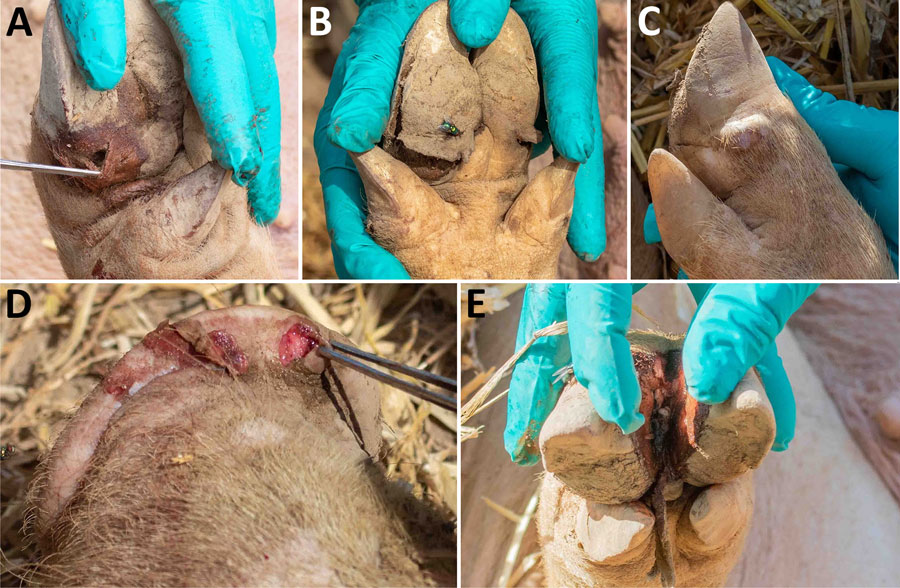
Figure 1. Affected pigs on farm SVV2022-02, from study of vesicular disease caused by Seneca Valley virus in pigs, England, 2022. Vesicular lesions can be seen on the coronary bands (A-C), snout (D), and interdigital cleft (E). Hoof horn separation also occurred in some infected pigs (B). Some lesions resembled those of foot-and-mouth disease (D), but others were more deep-seated (A).
Page created: February 15, 2026
Page updated: February 20, 2026
Page reviewed: February 20, 2026
The conclusions, findings, and opinions expressed by authors contributing to this journal do not necessarily reflect the official position of the U.S. Department of Health and Human Services, the Public Health Service, the Centers for Disease Control and Prevention, or the authors' affiliated institutions. Use of trade names is for identification only and does not imply endorsement by any of the groups named above.