Volume 32, Number 4—April 2026
Dispatch
Treatment of Severe Ocular Mpox with Cidofovir and Tecovirimat
Figure
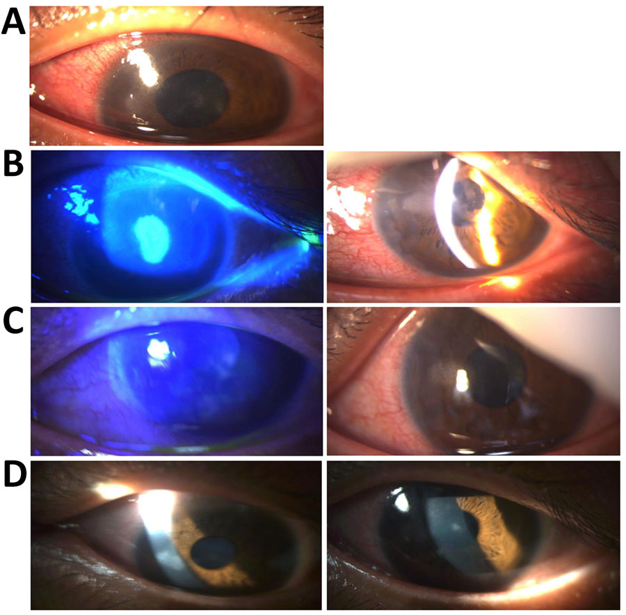
Figure. Ophthalmologic evaluation of patient with severe ocular mpox treated with cidofovir and tecovirimat. Images are of patient’s left eye over course of diagnosis and treatment. A) Slit-lamp examination at day 0, showing corneal edema, a pseudodendritic ulcer, and conjunctival hyperemia characteristic of disciform kerititis. B) On day 5, before first-line treatment with valaciclovir, fluorescein staining (left) and slit-lamp examination (right) showing persistent keratitis and formation of new corneal infiltrates. C) On day 21, seven days after initiation of second-line antiviral drug treatment with cidofovir and tecovirimat, fluorescein staining (left) and slit-lamp examination (right) showing marked clinical improvement and reduction in infiltrates. D) On day 29, fourteen days after treatment with second-line antiviral drug treatment with cidofovir and tecovirimat, left and right images are of slit-lamp examination showing complete resolution of corneal edema, conjunctival hyperemia, and inflammatory signs.