Volume 32, Number 6—June 2026
Dispatch
Suspected Sexual Transmission of Dermatophilosis among Men Who Have Sex with Men, Lyon and Paris, France, 2025–2026
Cite This Article
Citation for Media
Abstract
We report a genomically linked cluster of 9 Dermatophilus congolensis cutaneous infections diagnosed within 2 months among men who have sex with men in Lyon and Paris, France, 2025–2026. Genomic similarity and shared sexual exposures strongly suggest interhuman sexual transmission of this zoonotic bacterium.
Dermatophilus congolensis is a gram-positive, facultatively anaerobic actinomycete responsible for dermatophilosis, an exudative dermatitis of animals (1). The disease predominantly affects cattle, sheep, and horses, mainly in tropical and subtropical climates. It typically manifests as benign, crusting superficial skin lesions but occasionally progresses to extensive disease, sometimes resulting in significant mortality in cattle herds (2,3). The pathogenesis relies on 2 factors, skin microabrasions and moisture, which activate motile zoospores to penetrate the epidermis (1).
Human infections are rare and considered accidental zoonoses. Those infections are classically described in farmers, hunters, veterinarians, or animal riders following direct contact with infected animals (4–10). The clinical manifestation typically involves nonpruritic, pustular, and crusty lesions. D. congolensis is susceptible to β-lactams, macrolides, and tetracyclines. Systematic susceptibility testing is rarely performed in clinical practice. Reported treatments include penicillins, although lesions are often self-limiting. To date, human-to-human transmission has not been documented, and urban cases without reported animal exposure have rarely been described (11). We describe a temporally clustered series of human dermatophilosis cases occurring in France among urban men who have sex with men (MSM) without livestock exposure, raising the hypothesis of an alternative mode of transmission.
During December 2025–February 2026, a total of 9 men sought care at the sexually transmitted infections (STI) clinics of the University Hospital in Lyon, France, for skin infections that were determined to be caused by D. congolensis. All patients were MSM living in urban areas; they were 22–63 years of age (median 50, interquartile range [IQR] 34–59.5 years). None reported occupational exposure to livestock or direct contact with farm animals or horses, although some did report regular or occasional contact with domestic pets (cats or dogs). None reported recent travel to tropical regions.
Figure 1
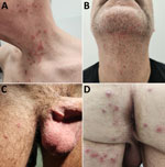
Figure 1. Dermatophilosis lesions in patients in cluster of suspected sexual transmission of dermatophilosis among men who have sex with men, Lyon and Paris, France, 2025–2026. A) Papular lesions of the neck...
All patients exhibited nonspecific erythematous papules, occasionally pustular or squamous, mainly located in the genital region (penis, scrotum, pubic area; n = 8), trunk (n = 5), perioral region (beard area; n = 4), lower limbs (n = 4), and, less frequently, anal margin (n = 1) (Table; Figure 1). Lesions predominantly involved areas exposed during sexual contact, without mucosal involvement. Pruritus was variably present. No systemic symptoms were reported except for patient 7, who experienced fever and vomiting 2 days before medical consultation. Given the clinical manifestations and epidemiologic context, differential diagnoses included staphylococcal folliculitis, sexually transmitted dermatophytosis (Trichophyton mentagrophytes genotype VII), Klebsiella aerogenes folliculitis, secondary syphilis, mpox, and molluscum contagiosum.
Figure 2
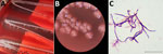
Figure 2. Results of sample testing in cluster of suspected sexual transmission of dermatophilosis among men who have sex with men, Lyon and Paris, France, 2025–2026. A, B) Dermatophilus congolensiscolonies...
For all patients, we cultured lesion swab samples from involved areas on nonselective media: blood agar plates (aerobic atmosphere) and chocolate agar plates (with 5% CO2). After 40 hours, cultures yielded β-hemolytic, rhizoid, adherent, rough, shiny, and yellowish colonies on both culture media (Figure 2, panels A, B), and occasionally along with colonies from the skin microbiota. Gram staining revealed identical filamentous, branching gram-positive bacilli with transverse and longitudinal septations producing a characteristic tram-track appearance (Figure 2, panel C). Bacterial identification by matrix-assisted laser desorption/ionization time-of-flight mass spectrometry identified D. congolensis with a high confidence score in all cases. In 4 patients, bacterial cultures also yielded pyogenic pathogens: Staphylococcus aureus (patients 3, 7 and 9) or S. lugdunensis (patient 8). Whole-genome sequencing (NextSeq550; Illumina, https://www.illumina.com) and pairwise alignment of D. congolensis isolates from patients 1–8 revealed 1–5 single-nucleotide polymorphisms, covering >94% of the genome, supporting close relatedness and recent direct or indirect transmission from a common source. Reads are available for consultation on the European Nucleotide Archive database (Table). Concomitant STIs were diagnosed in 2 patients (pubic pthiriasis in patient 1 and syphilitic reinfection in patient 7).
Epidemiologic interviews revealed that 7 of the 9 patients reported recent sexual encounters at a gay sauna in Lyon within days or weeks before symptom onset (Table). Patient 5 reported multiple sexual partners in various saunas in Paris during the same period, including 1 that patient 3 visited 2 days earlier. Patient 9 did not report any sauna attendance. On the basis of reported exposures, incubation period ranged from 3 to 14 days; survival modeling with interval censoring estimated a median of 6.7 (95% CI 4.3–10.1) days. However, multiple potential contacts limited precision (Table).
Amoxicillin MIC determined for 1 isolate using Etest (bioMérieux, https://www.biomerieux.com) was 0.064 mg/L, supporting high susceptibility to β-lactams. All patients received oral amoxicillin (1 g 3×/d) or pristinamycin (1 g 3×/d) for 7 days, sometimes combined with topical antiseptic care, with rapid improvement. No patient relapsed; median follow-up was 10 (IQR 5–52) days after treatment. However, on follow-up, patient 1 showed with a new D. congolensis infection on the buttocks, occurring 8 weeks after the first occurrence (Figure 1, panel D). His ongoing visits to the same sauna after recovery and the different infection site suggest reinfection rather than relapse.
Compared with previous reports of dermatophilosis, the predominantly papular and noncrusted manifestation observed here might differ from the classical description, raising the possibility of a distinct clinical phenotype. Moreover, lesions mainly affected areas exposed during sexual intercourse (face and genitals), mirroring patterns seen in sexually transmitted dermatophytosis or K. aerogenes folliculitis (12–14). Whereas no mucosal lesions were observed in this cluster, rare literature reports indicate that D. congolensis can infect human mucosa, justifying detailed clinical examination (6,11). In some occurrences, S. aureus and S. lugdunensis were also isolated from lesion sampling and might have modified clinical manifestations. Their involvement complicates the interpretation of D. congolensis pathogenicity in the given occurrences. Secondary infection of Dermatophilus-induced lesions by such pathogens is the most probable explanation, although co-transmission remains possible.
Although no direct sexual contact between patients could be formally established, the temporal clustering, overlapping sexual exposures, shared multiple STI history, lesion distribution, and close genomic relatedness of isolates strongly support transmission occurring within shared exposure networks, likely involving close physical or sexual contact. The presence of viable bacteria within lesions is consistent with the hypothesis of contact-driven transmission, although the exact transmission route remains uncertain. Environmental or indirect transmission within shared venues remains possible; no environmental sampling or carriage screening was performed at the time of the study to clarify transmission pathways.
We describe a large occurrence of human dermatophilosis cases in an urban population of sexually active MSM without reported livestock exposure. The combination of close genomic relatedness between the 8 sequenced isolates and shared sexual exposures suggest interhuman transmission within sexual networks. Systematic microbiologic evaluation of atypical cutaneous lesions is essential for identifying similar cases and clarifying transmission of emerging cutaneous STIs among MSM. Evolving sexual practices in the HIV preexposure prophylaxis era (15) could lead to emergence of new transmissible dermatoses (12–14). Microbiologists should ensure that D. congolensis is included in reference spectral libraries, be able to recognize its colonies in the context of skin infection, and report its identification to clinicians.
Dr. Degreze is a medical biology resident at the University Hospital in Lyon. Specializing in infectious agents, his research at the Center for Infectiology Research (CIRI) focuses on antistaphylococcal bacteriophages and the mechanisms of phage resistance. Alongside his research, he acts as a clinical microbiologist dedicated to the diagnosis of bacterial infections.
Acknowledgment
We are sincerely grateful to Didier Pin for his expertise on D. congolensis infections in animals and to Aubin Souche for genomic analysis and comparison.
References
- Zaria LT. Dermatophilus congolensis infection (Dermatophilosis) in animals and man! An update. Comp Immunol Microbiol Infect Dis. 1993;16:179–222. DOIPubMedGoogle Scholar
- Faccin M, Wiener DJ, Rech RR, Santoro D, Rodrigues Hoffmann A. Common superficial and deep cutaneous bacterial infections in domestic animals: a review. Vet Pathol. 2023;60:796–811. DOIPubMedGoogle Scholar
- Naves M, Vallée F, Barré N. Observations on a dermatophilosis outbreak in Brahman cattle in Guadeloupe. Description, epidemiological and economical aspects [in French]. Rev Elev Med Vet Pays Trop. 1993;46:297–302. DOIPubMedGoogle Scholar
- Burd EM, Juzych LA, Rudrik JT, Habib F. Pustular dermatitis caused by Dermatophilus congolensis. J Clin Microbiol. 2007;45:1655–8. DOIPubMedGoogle Scholar
- Amor A, Enríquez A, Corcuera MT, Toro C, Herrero D, Baquero M. Is infection by Dermatophilus congolensis underdiagnosed? J Clin Microbiol. 2011;49:449–51. DOIPubMedGoogle Scholar
- Bunker ML, Chewning L, Wang SE, Gordon MA. Dermatophilus congolensis and “hairy” leukoplakia. Am J Clin Pathol. 1988;89:683–7. DOIPubMedGoogle Scholar
- Aubin GG, Guillouzouic A, Chamoux C, Lepelletier D, Barbarot S, Corvec S. Two family members with skin infection due to Dermatophilus congolensis: a case report and literature review. Eur J Dermatol. 2016;26:621–2. DOIPubMedGoogle Scholar
- de Lorenzi C, Quenan S, Fontao L. Dermatophilus congolensis dermatitis in a traveller from Thailand. J Travel Med. 2021;28:
taab017 . DOIPubMedGoogle Scholar - Alejo-Cancho I, Bosch J, Vergara A, Mascaro JM, Marco F, Vila J. Dermatitis by Dermatophilus congolensis. Clin Microbiol Infect. 2015;21:e73–4. DOIPubMedGoogle Scholar
- Towersey L, Martins EC, Londero AT, Hay RJ, Soares Filho PJ, Takiya CM, et al. Dermatophilus congolensis human infection. J Am Acad Dermatol. 1993;29:351–4. DOIPubMedGoogle Scholar
- Ramanathan VS, Jahng AW, Shlopov B, Pham BV. Dermatophilus congolensis infection of the esophagus. Gastroenterol Res. 2010;3:173–4. DOIPubMedGoogle Scholar
- Jabet A, Dellière S, Seang S, Chermak A, Schneider L, Chiarabini T, et al. Sexually transmitted Trichophyton mentagrophytes genotype VII infection among men who have sex with men. Emerg Infect Dis. 2023;29:1411–4. DOIPubMedGoogle Scholar
- Bérot V, Monsel G, Dauendorffer JN, Aubry A, Nebbad B, Schneider P, et al.; Groupe Infectiologie Dermatologique et Infections Sexuellement Transmissibles. (GrIDIST) de la Société Française de Dermatologie. Klebsiella aerogenes–related facial folliculitis in men having sex with men: a hypothetical new STI? J Eur Acad Dermatol Venereol. 2025;39:e10–2. DOIPubMedGoogle Scholar
- Yin N, Krygier J, Mairesse C, Libois A, Quoilin S, Martiny D. Klebsiella aerogenes ST117 causing folliculitis in men having sex with men, Belgium, February 2025. Euro Surveill. 2025;30:
2500304 . DOIPubMedGoogle Scholar - Milam J, Jain S, Dubé MP, Daar ES, Sun X, Corado K, et al. CCTG Team. Sexual risk compensation in a pre-exposure prophylaxis demonstration study among individuals at risk of HIV. J Acquir Immune Defic Syndr. 2019;80:e9–13. DOIPubMedGoogle Scholar
Figures
Table
Cite This ArticleOriginal Publication Date: April 30, 2026
Table of Contents – Volume 32, Number 6—June 2026
| EID Search Options |
|---|
|
|
|
|
|
|
Please use the form below to submit correspondence to the authors or contact them at the following address:
Matthieu Degreze, Service de bactériologie, Institut des Agents Infectieux, Laboratoire de Biologie Médicale Multisites, Hospices Civils de Lyon, 103 Grande Rue de la Croix-Rousse, 69004 Lyon, France
Top