Volume 32, Number 6—June 2026
Dispatch
Suspected Sexual Transmission of Dermatophilosis among Men Who Have Sex with Men, Barcelona, Spain, 2025–2026
Cite This Article
Citation for Media
Abstract
Dermatophilosis is considered a zoonotic infection. We report 9 cases among men who have sex with men in Barcelona, Spain, during December 2025–March 2026. Whole-genome sequencing revealed highly related isolates forming a distinct lineage within the genus Dermatophilus. Epidemiologic and clinical features of the cases support human-to-human transmission via sexual contact.
Dermatophilosis is a skin infection caused by Dermatophilus congolensis, a gram-positive actimoycete that primarily affects animals in tropical and subtropical regions (1). Human infections are rare and typically associated with zoonotic exposure to livestock or wildlife; manifestations include pitted keratolysis, folliculitis, and pustules (2–6). Human-to-human transmission has not been documented. Here, we report 9 cases of dermatophilosis diagnosed in Barcelona, Spain, and assess the possibility of autochthonous human-to-human transmission via sexual contact.
The cases involved 2 patients who attended primary healthcare centers in December 2025 and March 2026 and 7 patients who attended a reference clinic for sexually transmitted infections (STIs) during January–March 2026 (Table). All patients were cisgender men who reported having sex with men (MSM). The median age was 47 years (interquartile range 30.5–57.5 years). Four patients were HIV-positive and 3 were receiving HIV preexposure prophylaxis therapy. Four patients had concomitant STIs, and 3 reported engaging in chemsex (drug use in a sexual context).
None of the patients reported contact with livestock or wildlife, and none had traveled to tropical regions in the month before symptom onset. Four patients reported recent travel to other cities in Europe where they engaged in sexual activity. All patients reported visiting venues for sexual encounters within the week preceding symptom onset, including 8 who had visited a sauna. Two patients were regular sexual partners, and 2 others reported partners with similar symptoms who were treated in other centers without microbiological confirmation.
Figure 1

Figure 1. Dermatophilosis lesions in patients in cluster of suspected sexual transmission among men who have sex with men, Barcelona, Spain, December 2025–March 2026. A) Papule-pustules on the beard area; B) papulovesicular...
Median symptom duration at consultation was 6 days (interquartile range 4.5–15 days). Lesions manifested as an itchy folliculitis-like rash characterized by papules, vesicles, pustules, scabs, nodules, or scaly lesions (Figure 1). Multiple anatomic sites were affected, most commonly the genitals, thighs, groin, and beard area.
Five patients were treated with cefadroxil (500 mg 2×/d for 7 d) resulting in complete resolution of lesions in all but 1 case, who had a residual nodular lesion. One patient was successfully treated with cloxacillin (500 mg/6 h for 7 d). Two patients showed cleared lesions after treatment for concomitant STIs (benzathine penicillin G for syphilis and ceftriaxone and doxycycline for gonorrhea and lymphogranuloma venereum). One patient initially received topical mupirocin with partial response but achieved complete resolution after doxycycline therapy (100 mg 2×/d for 7 d). None of the patients required hospitalization or experienced any complications. As of April 2026, no recurrences had been observed.
Skin lesion samples cultured on Columbia 5% sheep blood agar produced small yellowish β-hemolytic colonies. Gram staining revealed gram-positive coccoid forms occasionally arranged in irregular filaments with transverse septa. Identification by matrix-assisted laser desorption/ionization time-of-flight mass spectrometry confirmed Dermatophilus congolensis.
All isolates showed identical antimicrobial susceptibility profiles and were susceptible to amoxicillin/clavulanate (MIC <2/1 mg/L), ceftriaxone (MIC <4 mg/L), cefepime (MIC <1 mg/L), imipenem (MIC <2 mg/L), linezolid (MIC 1 mg/L), amikacin (MIC 4 mg/L), moxifloxacin (MIC <0.25 mg/L), doxycycline (MIC <0.12 mg/L), minocycline (MIC <1 mg/L), clarithromycin (MIC <0.06 mg/L), and trimethoprim/sulfamethoxazole (MIC <0.25/4.75 mg/L). All isolates were resistant to tobramycin (MIC 16 mg/L) and showed intermediate susceptibility to ciprofloxacin (MIC 2 mg/L). We interpreted susceptibility on the basis of CLSI 2023 breakpoints for other aerobic actinomycetes (7) because no organism-specific breakpoints are available for D. congolensis. MIC for mupirocin was >1,024 mg/L for all isolates; no clinical breakpoints are available.
Figure 2
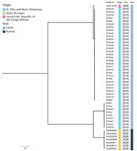
Figure 2. Phylogenetic analysis of Dermatophilus isolates in cluster of suspected sexual transmission of dermatophilosis among men who have sex with men, Barcelona, Spain, December 2025–March 2026. Maximum-likelihood phylogenetic tree shows ...
We performed whole-genome sequencing (WGS) on isolates from patients 1–7. WGS demonstrated extremely close genetic relatedness among isolates; pairwise single-nucleotide polymorphism (SNP) distances were 0–4 SNPs. We also performed phylogenetic comparisons between the isolates from this study and publicly available D. congolensis genomes, including 40 genomes from cattle in Saint Kitts and Nevis (8) and reference strain D. congolensis DSM 44180 from the Democratic Republic of the Congo, all from the National Center for Biotechnology Information Sequence Read Archive (https://www.ncbi.nlm.nih.gov/sra). Those comparisons showed that the study isolates formed a distinct cluster clearly separated from previously described strains; minimum SNP distance was 20,410 to the closest reference genome (Figure 2).
Genomic comparison with reference strain D. congolensis DSM 44180 yielded an average nucleotide identity of 94.55%, below the species delineation threshold of 95%–96% (9). Digital DNA–DNA hybridization values among study isolates were 99.9%–100%, whereas values compared with the reference strain were 58.7%, below the 70% threshold for species delineation (9). Those findings suggest that the isolates are genetically distinct from currently described D. congolensis strains and are consistent with a potentially novel Dermatophilus taxon, although further taxonomic characterization would be required to formally define a new species.
We report human cases of dermatophilosis for which no animal exposure was identified and which are suspected to have been sexually acquired. All cases occurred in MSM with high exposure to STIs, several patients reported partners with similar symptoms, and lesions were commonly located in sites exposed during sexual contact. Those features were similarly described for other considered zoonotic pathogens that emerged as sexually transmissible, such as mpox virus (10) and Trichophyton mentagrophytes (11). As has already been observed, MSM are particularly vulnerable to such emerging infections, probably because of the complexity of their sexual networks (12).
Attendance at sexual venues might have been a factor in transmission in this cluster. In particular, 8 patients developed lesions shortly after visiting a sauna, where humid conditions could favor the release and environmental persistence of infective Dermatophilus zoospores (13,14). Indirect transmission might occur via contaminated surfaces; fomite-mediated outbreaks have been described in animals (15). However, based on the anatomic distribution of lesions, direct skin-to-skin contact during sexual activity likely represents the main route of transmission.
All cases in this series were mild and resolved easily without complications. Most patients responded well to short courses of commonly used antibiotics, including β-lactams or doxycycline. That finding is consistent with previous reports of human dermatophilosis as a mostly benign condition (2–6). Although dermatophilosis might resolve spontaneously over several weeks, accelerated recovery through antibiotic therapy could help reduce transmission within the community. Antimicrobial susceptibility testing showed a broad susceptibility profile to several antibiotic classes, suggesting that standard oral treatments remain effective. In the case of mupirocin, the finding of a high MIC suggests that this topical antibiotic might not be the most suitable treatment option.
Genomic findings support recent Dermatophilus transmission among humans. The WGS of isolates showed a close genetic relationship, consistent with a recent common ancestor and short transmission chains. The isolates formed a well-supported phylogenetic cluster distinct from publicly available D. congolensis genomes, suggesting the circulation of a single lineage rather than multiple independent introductions and representing a novel taxon within the genus Dermatophilus. This previously undescribed taxon circulating in humans could potentially contribute to the epidemiologic pattern observed, although further studies are needed to clarify its ecologic niche, host range, and transmission dynamics.
In summary, this cluster of genetically closely related cases of dermatophilosis within sexual networks suggests that this condition might be emerging as a sexually transmissible infection, although environmental transmission cannot be excluded. Because clinical manifestations can be nonspecific and laboratory identification is uncommon in STI clinics, cases could remain unrecognized. Clinicians should therefore suspect dermatophilosis in MSM who have a folliculitis-like pustular rash involving genital or adjacent areas and should consider oral antibiotic treatment and comprehensive STI screening. Cross-border surveillance could help determine whether similar cases are occurring elsewhere.
Dr. Descalzo is an internal medicine specialist working in the STI/HIV unit Drassanes-Hospital Vall d’Hebron in Barcelona, Spain. His primary research interest is in emerging sexually transmissible infections.
Acknowledgments
This work was carried out within the frame of the Doctoral Program in Medicine at the Universitat Autonoma de Barcelona. The study was approved by the Ethics Committee of Vall d’Hebron Hospital, reference no. PR(AG)080/2026. The study was conducted in accordance with the principles laid out in the Declaration of Helsinki and in accordance with the principles of Good Clinical Practice. Informed consent was obtained from participants prior to the inclusion of their data in the analysis, including authorization for the publication of clinical images.
WGS data generated in this study has been deposited in the National Center for Biotechnology Information Sequence Read Archive under BioProject accession no. PRJNA1433182. All D. congolensis sequences used in this study, including accession numbers and associated metadata, are described in the Appendix.
This study was partially supported by the “Centro de Investigación Biomédica en Red” (CIBER de Enfermedades Infecciosas; grant no. CB21/13/00054).
References
- Zaria LT. Dermatophilus congolensis infection (Dermatophilosis) in animals and man! An update. Comp Immunol Microbiol Infect Dis. 1993;16:179–222. DOIPubMedGoogle Scholar
- Dean DJ, Gordon MA, Severinghaus CW, Kroll E, Reilly JR. Streptothricosis: a new zoonotic disease. N Y State J Med. 1961;61:1285–7.
- Towersey L, Martins EC, Londero AT, Hay RJ, Soares Filho PJ, Takiya CM, et al. Dermatophilus congolensis human infection. J Am Acad Dermatol. 1993;29:351–4. DOIPubMedGoogle Scholar
- Burd EM, Juzych LA, Rudrik JT, Habib F. Pustular dermatitis caused by Dermatophilus congolensis. J Clin Microbiol. 2007;45:1655–8. DOIPubMedGoogle Scholar
- Alejo-Cancho I, Bosch J, Vergara A, Mascaro JM, Marco F, Vila J. Dermatitis by Dermatophilus congolensis. Clin Microbiol Infect. 2015;21:e73–4. DOIPubMedGoogle Scholar
- Aubin GG, Guillouzouic A, Chamoux C, Lepelletier D, Barbarot S, Corvec S. Two family members with skin infection due to Dermatophilus congolensis: a case report and literature review. Eur J Dermatol. 2016;26:621–2. DOIPubMedGoogle Scholar
- Clinical and Laboratory Standards Institute. Performance standards for susceptibility testing of mycobacteria, Nocardia spp., and other aerobic actinomycetes (document M24S). Wayne (PA): The Institute; 2023.
- Branford I, Boyen F, Johnson S, Zayas S, Chapwanya A, Butaye P, et al. Identification and antimicrobial resistance of Dermatophilus congolensis from cattle in Saint Kitts and Nevis. Vet Sci. 2021;8:135. DOIPubMedGoogle Scholar
- Riesco R, Trujillo ME. Update on the proposed minimal standards for the use of genome data for the taxonomy of prokaryotes. Int J Syst Evol Microbiol. 2024;74:
006300 . DOIPubMedGoogle Scholar - Tarín-Vicente EJ, Alemany A, Agud-Dios M, Ubals M, Suñer C, Antón A, et al. Clinical presentation and virological assessment of confirmed human monkeypox virus cases in Spain: a prospective observational cohort study. Lancet. 2022;400:661–9. DOIPubMedGoogle Scholar
- Descalzo V, Martín MT, Álvarez-López P, García-Pérez JN, Alcázar-Fuoli L, López-Pérez L, et al. Trichophyton mentagrophytes genotype VII and sexually transmitted tinea: an observational study in Spain. Mycoses. 2025;68:
e70049 . DOIPubMedGoogle Scholar - Spicknall IH, Gift TL, Bernstein KT, Aral SO. Sexual networks and infection transmission networks among men who have sex with men as causes of disparity and targets of prevention. Sex Transm Infect. 2017;93:307–8. DOIPubMedGoogle Scholar
- Abu-Samra MT. The epizootiology of Dermatophilus congolensis infection. Rev Elev Med Vet Pays Trop. 1980;33:23–32.PubMedGoogle Scholar
- Martinez D, Prior P. Survival of Dermatophilus congolensis in tropical clay soils submitted to different water potentials. Vet Microbiol. 1991;29:135–45. DOIPubMedGoogle Scholar
- García Sánchez A, Zurita SG, Gil Molino M, Martin Cano FE, Barraso Gil C, Hermoso de Mendoza Salcedo J. Outbreak of dermatophilosis in horses possibly transmitted by sharing riding equipment. Braz J Vet Med. 2024;46:
e002124 . DOIPubMedGoogle Scholar
Figures
Table
Cite This ArticleOriginal Publication Date: April 30, 2026
1These first authors contributed equally to this article.
2These senior authors contributed equally to this article.
Table of Contents – Volume 32, Number 6—June 2026
| EID Search Options |
|---|
|
|
|
|
|
|
Please use the form below to submit correspondence to the authors or contact them at the following address:
Maider Arando, STI/HIV Unit Drassanes–Vall d’Hebron, Hospital Universitari Vall d’Hebron, 17 Sant Oleguer, 08001 Barcelona, Spain
Top