Disclaimer: Early release articles are not considered as final versions. Any changes will be reflected in the online version in the month the article is officially released.
Volume 32, Number 7—July 2026
Dispatch
Nipah Virus Shedding in Urine from Fruit Bats, Sri Lanka, 2018–2019
Suggested citation for this article
Abstract
Nipah virus causes outbreaks in humans with high case-fatality rates. In this study, we confirmed the presence of Nipah virus in Sri Lanka in Pteropus medius fruit bats, one of the known natural reservoir species. Sequences we generated were genetically related to Nipah virus strains from outbreaks in southern India.
Nipah virus (NiV; Henipavirus nipahense) and Hendra virus (H. hendraense) are species within the genus Henipavirus of the family Paramyxoviridae (1). Several paramyxoviruses, including measles virus and respiratory syncytial virus, cause serious respiratory illnesses and are often highly transmissible through the air. NiV can cause severe outbreaks in humans; its clinical symptoms range from subclinical infection to severe encephalitis, respiratory diseases, and death (2). Reported case-fatality rate (CFR) of NiV encephalitis is 61% (95% CI 45.7%–75.4%) (2,3).
NiV emerged in 1998 in Malaysia and Singapore (4). Subsequent outbreaks were reported from Bangladesh, the Philippines, and India; they resulted in >643 laboratory-confirmed infections and 380 deaths (2). Bats (flying foxes, Pteropus spp.) are the known reservoir hosts of NiV (5). Transmission from bat to human occurs via exposure to urine, either through consumption of raw palm sap contaminated with bat excreta, direct contact with infected intermediate hosts (e.g., swine), or direct contact with bat urine (5). In 2023 several cases of Nipah virus were reported from Kozhikode, Kerala district, India, and in Dhaka, Rajbari, and Shariatpur districts in Bangladesh (6). The shortest distance between Mannar Island, Sri Lanka, and Natarajapuram, India, is <55 km; Pteropus spp. bats are reported to migrate >450 km with ease (7). In this study, we monitored urine excreted by several colonies of P. medius bats in Sri Lanka for the presence of viruses using molecular techniques. Our focus was to investigate if NiV is present in Sri Lanka and to determine the measures required to prevent spillover into humans. We obtained ethics approval for this study from the Institute of Biology, Sri Lanka (WL/3/2/05/18) and necessary clearance from the Department of Wildlife Conservation, Sri Lanka.
During March 2018–June 2019, we monitored P. medius bat colonies for shedding of pathogens in Colombo, Mannar, Anuradhapura, and Badulla, Sri Lanka. We laid clean sampling sheets below roosting trees in the early morning and transferred excreted urine drop by drop to sterile microtiter plates in the morning using disposable pipettes. We conducted all work while wearing appropriate personal protective equipment (PPE) as previously described (5).
We collected a total of 2,218 urine samples and combined them into 32 pools with <8 urine specimens each. We extracted RNA from the individual pools using the QIAamp Viral RNA Mini Kit (QIAGEN, https://www.qiagen.com). We performed Nipah virus screening using real-time PCR targeting the phosphoprotein (P) gene, as previously described (8). As an additional confirmation, we developed a real-time PCR targeting the nucleocapsid protein of the Nipah virus genome (Table 1).
Figure 1
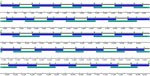
Figure 1. Positions of primers used for NiVliSeq amplification panel in study of Nipah virus shedding in urine from fruit bats, Sri Lanka, 2018–2019. Blue and green represent 2 nonoverlapping primer pools...
We used Illumina HiSeq (https://www.illumina.com) to sequence the NiV positive pools S3_P02 and S18_P08 (2 of 32 pools). We trimmed using Trimmomatic (9) and mapped to NiV strain MCL-18-H-1088 (accession no. MHK523642; Kerala, India) as a reference strain. In addition, we developed an AmpliSeq primer panel, NiVliSeq, spanning the whole NiV genome using Primer3 version 2.3.7 (Figure 1; Appendix Table). We sequenced corresponding PCR products on the MinIon platform as described previously (10,11). We submitted draft genomes to GenBank (accession nos. PP893186–9) and aligned them with NiV strains and type species of paramyxovirus genomes indicated by the International Committee on Taxonomy of Viruses and available in GenBank. We calculated phylogenetic molecular clock reconstruction with MrBayes version 0.39 software (https://nbisweden.github.io/MrBayes) using Markov chain Monte Carlo approach of molecular clock reconstruction based on the general time reversible model with the parameters set at 1 million replicates, 4 chains, and burn in at 10% (partial large sequence 7,977 nt; coding for the RNA-dependent RNA polymerase gene, of the subfamily Orthoparamyxovirinae).
We collected pool S3_P02 and pool S18_P08 in Colombo; both tested positive for NiV using the henipavirus quantitative PCR (qPCR) targeting the P gene (8). The NiV qPCR (targeting the nucleocapsid gene) further confirmed NiV in the same sample pools (Table 2). Sequencing revealed corresponding NiV reads in the 2 sample pools that we mapped over the entire NiV genome (Table 2), providing additional confirmation. The NiVliSeq approach revealed 93% of the NiV genome from pool S3_P02 and 98% of NiV genome for pool S18-08, (Table 2). Comparison of the NiV genomes with the GenBank database revealed the highest identity to a strain collected from a human patient during the 2018 outbreak in Kerala, India (accession no. MH396625); the genomes from pool S3_P02 and pool S18-08 each had 98% nucleotide identity to the Kerala strain. We named the Sri Lanka NiV strains in accordance with the system used for the India strains: NiV strain C-18-B-0302 Sri Lanka (accession no. PP893186.1) and NiV strain C-19-B-1808 Sri Lanka (accession no. PP893188.1). In that nomenclature, C denotes Colombo, 18 and 19 indicate the years 2018 and 2019, B denotes bat, and the remaining numbers represent the sample number.
Figure 2
Figure 2. Phylogenetic reconstruction of subfamily Orthoparamyxovirinae viruses in study of NiV shedding in urine from fruit bats, Sri Lanka, 2018–2019. The alignment is based on the partial large gene (7,977 nt)...
Phylogenetic analysis allocated the novel Nipah virus strains C-18-B-0302 Sri Lanka and C-19-B-1808 Sri Lanka to the species Nipah virus (H. nipahense) within the genus Henipavirus (Figure 2). Both are clustering monophyletically with the strains sampled from humans during the outbreaks in Kerala region, India in 2007, 2018, 2019, 2021, and 2023.
This study demonstrated the presence of NiV strains in Sri Lanka. The qPCR and NiVliSeq sequencing approach we developed validated previously obtained results and represented valuable tools that can be used in further studies. The identified strains have highest similarities to human-pathogenic strains causing recent outbreaks in India (2007, 2018, 2019, and 2023). However, NiV is still a rare disease, and transmission rates are comparably low. Furthermore, the detection of a certain virus in a bat population does not necessarily mean that transmission occurs; diverse strains of henipaviruses can be found in Pteropus spp. bats across the whole distribution range.
The initial prevention of any infection should be one of the major goals and could include protection of the bats and their roosts. Bat colonies are vital for healthy ecosystems and should be protected for several reasons, including the ecosystem services they provide such as pollination and seed dispersal. Moreover, Pteropus spp. bats are threatened by extinction (12). The World Health Organization (WHO) has recognized the need for research and diagnosis of NiV within the distribution range of Pteropus spp. (13).
Understanding the mechanisms influencing shedding of NiV will likely prevent further spread; NiV shedding likely also follows seasonal and temporal pulses previously reported for Hendra virus (14–15). We recognize that disease emergence might be a result of disturbance within ecologic systems and hence cannot be alleviated by further disturbance. As a tropical country, Sri Lanka has complex and diverse ecosystems; its resilience to climate change may be better than other regions of the world.
The potential of zoonotic bat-to-human transmission of NiV could be minimized by preventive measures. Such measures could include education of healthcare workers to raise appropriate awareness for NiV, equipment and training for the early detection of NiV RNA in clinical specimens, preparation for clinical management of NiV-positive patients, and messaging the public about avoidance of known infection routes to prevent transmission (i.e., circular fencing of trees with roosting bats).
Dr. Kohl is a scientist at the Robert Koch Institute in Berlin, Germany, at the Centre for Biological Threats and Special Pathogens. Her research interests are the detection of emerging and reemerging viruses and the characterization of novel pathogens, especially from bats.
Acknowledgment
This article was preprinted at https://www.biorxiv.org/content/10.1101/2024.07.31.605971v1
References
- Rima B, Balkema-Buschmann A, Dundon WG, Duprex P, Easton A, Fouchier R, et al.; ICTV Report Consortium. ICTV virus taxonomy profile: Paramyxoviridae. J Gen Virol. 2019;100:1593–4. DOIPubMedGoogle Scholar
- Sharma V, Kaushik S, Kumar R, Yadav JP, Kaushik S. Emerging trends of Nipah virus: a review. Rev Med Virol. 2019;29:
e2010 . DOIPubMedGoogle Scholar - Kenmoe S, Demanou M, Bigna JJ, Nde Kengne C, Fatawou Modiyinji A, Simo FBN, et al. Case fatality rate and risk factors for Nipah virus encephalitis: A systematic review and meta-analysis. J Clin Virol. 2019;117:19–26. DOIPubMedGoogle Scholar
- Chua KB, Goh KJ, Wong KT, Kamarulzaman A, Tan PS, Ksiazek TG, et al. Fatal encephalitis due to Nipah virus among pig-farmers in Malaysia. Lancet. 1999;354:1257–9. DOIPubMedGoogle Scholar
- Chua KB. A novel approach for collecting samples from fruit bats for isolation of infectious agents. Microbes Infect. 2003;5:487–90. DOIPubMedGoogle Scholar
- As AK, Sahay RR, Radhakrishnan CPS, Kandath S, Patil DY, et al. Clinico-epidemiological presentations and management of Nipah virus infection during the outbreak in Kozhikode district, Kerala state, India 2023. J Med Virol. 2024;96:
e29559 . DOIPubMedGoogle Scholar - Roberts BJ, Catterall CP, Eby P, Kanowski J. Long-distance and frequent movements of the flying-fox Pteropus poliocephalus: implications for management. PLoS One. 2012;7:
e42532 . DOIPubMedGoogle Scholar - Feldman KS, Foord A, Heine HG, Smith IL, Boyd V, Marsh GA, et al. Design and evaluation of consensus PCR assays for henipaviruses. J Virol Methods. 2009;161:52–7. DOIPubMedGoogle Scholar
- Bolger AM, Lohse M, Usadel B. Trimmomatic: a flexible trimmer for Illumina sequence data. Bioinformatics. 2014;30:2114–20. DOIPubMedGoogle Scholar
- Brinkmann A, Ulm SL, Uddin S, Förster S, Seifert D, Oehme R, et al. AmpliCoV: rapid whole-genome sequencing using multiplex PCR amplification and real-time Oxford Nanopore MinION sequencing enables rapid variant identification of SARS-CoV-2. Front Microbiol. 2021;12:
651151 . DOIPubMedGoogle Scholar - Brinkmann A, Pape K, Uddin S, Woelk N, Förster S, Jessen H, et al. Genome sequencing of the mpox virus 2022 outbreak with amplicon-based Oxford Nanopore MinION sequencing. J Virol Methods. 2024;325:
114888 . DOIPubMedGoogle Scholar - International Union for Conservation of Nature and Natural Resources. IUCN Red List: Indian flying fox. 2021 [cited 2019 Aug 14]. DOIGoogle Scholar
- World Health Organization. WHO South-East Asia Regional Strategy for the prevention and control of Nipah virus infection 2023–2030. 2023 [cited 2024 Feb 3] https://www.who.int/publications/i/item/9789290210849
- Plowright RK, Peel AJ, Streicker DG, Gilbert AT, McCallum H, Wood J, et al. Transmission or within-host dynamics driving pulses of zoonotic viruses in reservoir-host populations. PLoS Negl Trop Dis. 2016;10:
e0004796 . DOIPubMedGoogle Scholar - Plowright RK, Eby P, Hudson PJ, Smith IL, Westcott D, Bryden WL, et al. Ecological dynamics of emerging bat virus spillover. Proc Biol Sci. 2015;282:
20142124 . DOIPubMedGoogle Scholar
Figures
Tables
Suggested citation for this article: Kohl C, Siriwardana S, Muzeniek T, Perera T, Bas D, Öruc M, et al. Nipah virus shedding in urine from fruit bats, Sri Lanka, 2018–2019. Emerg Infect Dis. 2026 Jul [date cited]. https://doi.org/10.3201/eid3207.251567
Original Publication Date: June 17, 2026
1These first authors contributed equally to this article.
Table of Contents – Volume 32, Number 7—July 2026
| EID Search Options |
|---|
|
|
|
|
|
|
Please use the form below to submit correspondence to the authors or contact them at the following address:
Claudia Kohl, Centre for Biological Threats and Special Pathogens, Robert Koch Institute, Seestraße 10, 13352 Berlin, Germany
Top