Volume 32, Number 7—July 2026
Research Letter
Autochthonous Neurocysticercosis Brain Lesions Mimicking Metastatic Disease, Spain
Figure
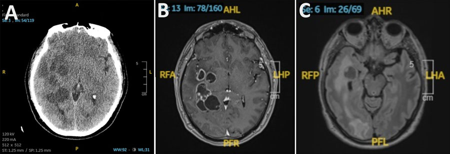
Figure. Radiologic findings from a study of autochthonous neurocysticercosis brain lesions mimicking metastatic disease, Spain. A) Noncontrast head computed tomography demonstrating multiple intra-axial lesions with surrounding vasogenic edema. B) Axial T1-weighted magnetic resonance imaging sequence with gadolinium showing multiple ring-enhancing lesions. C) Axial T2-FLAIR magnetic resonance imaging sequence revealing cystic lesions with internal nodular components suggestive of a scolex, surrounded by extensive edema.
Page created: June 08, 2026
Page updated: June 26, 2026
Page reviewed: June 26, 2026
The conclusions, findings, and opinions expressed by authors contributing to this journal do not necessarily reflect the official position of the U.S. Department of Health and Human Services, the Public Health Service, the Centers for Disease Control and Prevention, or the authors' affiliated institutions. Use of trade names is for identification only and does not imply endorsement by any of the groups named above.