Skip directly to site content Skip directly to page options Skip directly to A-Z link Skip directly to A-Z link Skip directly to A-Z link

Disclaimer: Early release articles are not considered as final versions. Any changes will be reflected in the online version in the month the article is officially released.

Volume 32, Number 8—August 2026

Research Letter

Human Lactococcus garvieae Bloodstream Infection Complicated by Spondylodiscitis, Germany

Philipp SchulzComments to Author , Pascal Migaud, Daniela Drauz, Sebastian Jaeger, Johannes Elias, and Hartmut Stocker
Author affiliation: Joseph-Kliniken–St. Joseph Krankenhaus Berlin–Tempelhof, Berlin, Germany (P. Schulz, P. Migaud, D. Drauz, S. Jaeger, H. Stocker); Charité–Universitätsmedizin Berlin, Berlin (P. Schulz, P. Migaud, H. Stocker); MDI Limbach Berlin GmbH, Berlin (J. Elias); Health and Medical University Potsdam, Potsdam, Germany (J. Elias)

Main Article

Figure 1

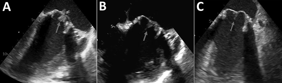
Transesophageal echocardiograph findings from a case of human Lactococcus garvieae bloodstream infection complicated by spondylodiscitis, Germany. A) Echocardiograph obtained on day 6 demonstrates mobile vegetations on both leaflets of the biological prosthetic mitral valve with a maximum dimension of 6 × 16 mm and pronounced valvular leaflet thickening (arrow). No notable valvular dysfunction was present. B) Echocardiograph obtained on day 45 shows marked regression of mitral valve leaflet thickening and a smaller vegetation (arrow; maximum 4 × 4 mm). No notable valvular dysfunction was present. C) Echocardiograph obtained on day 98 shows marked regression of mitral valve leaflet thickening. Arrow shows a faint 5 mm filamentous structure that might represent a residual finding. No notable valvular dysfunction was present.

Figure 1. Transesophageal echocardiograph findings from a case of human Lactococcus garvieae bloodstream infection complicated by spondylodiscitis, Germany. A) Echocardiograph obtained on day 6 demonstrates mobile vegetations on both leaflets of the biological prosthetic mitral valve with a maximum dimension of 6 × 16 mm and pronounced valvular leaflet thickening (arrow). No notable valvular dysfunction was present. B) Echocardiograph obtained on day 45 shows marked regression of mitral valve leaflet thickening and a smaller vegetation (arrow; maximum 4 × 4 mm). No notable valvular dysfunction was present. C) Echocardiograph obtained on day 98 shows marked regression of mitral valve leaflet thickening. Arrow shows a faint 5 mm filamentous structure that might represent a residual finding. No notable valvular dysfunction was present.

Main Article

Page created: June 23, 2026
Page updated: July 17, 2026
Page reviewed: July 17, 2026
The conclusions, findings, and opinions expressed by authors contributing to this journal do not necessarily reflect the official position of the U.S. Department of Health and Human Services, the Public Health Service, the Centers for Disease Control and Prevention, or the authors' affiliated institutions. Use of trade names is for identification only and does not imply endorsement by any of the groups named above.
file_external