Volume 6, Number 5—October 2000
Research
Imported Lassa Fever in Germany: Molecular Characterization of a New Lassa Virus Strain
Figure 3
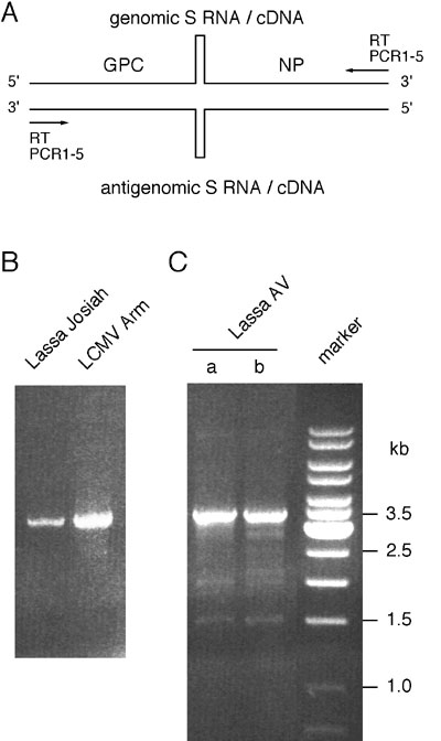
Figure 3. Reverse transcription (RT) and polymerase chain reaction (PCR) amplification of full-length S RNA. (A) Position of the RT and PCR primers at the termini of S RNA. The stem-loop structure in the intergenic region is schematically shown. (B) Amplified S RNA of Lassa Josiah and LCMV Armstrong virus separated in ethidium bromide-stained agarose gel. S RNA was isolated from supernatant of infected cells, and PCR was done with primers PCR2-4. (C) S RNA of Lassa AV was amplified in two RT-PCRs (a and b) and separated in ethidium bromide-stained agarose gel. RNA was isolated from serum, and PCR was performed with primers PCR2-4. Quantification of Lassa virus RNA in the specimen by endpoint titration with the 340-bp PCR assay showed >106 S RNA molecules/mL serum.