Volume 7, Number 4—August 2001
THEME ISSUE
West Nile Virus
West Nile Virus
West Nile Virus Infection in the Golden Hamster (Mesocricetus auratus): A Model for West Nile Encephalitis
Shu-Yuan Xiao, Hilda Guzman, Hui Zhang, Amelia P.A. Travassos da Rosa, and Robert B. Tesh
Figure 3
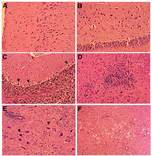
Figure 3. . Histologic changes in brains of West Nile virus-infected hamsters. A. Cerebral cortex, with many degenerating neurons, day 6 postinfection. B. Hippocampus, showing large neurons undergoing degeneration, day 6. C. Cerebellar cortex, with frequent Purkinje call degeneration (shrunken cells, arrowheads) and loss, day 8. D. A microglial nodule near blood vessel in basal ganglia, day 9. E. Mild perivascular inflammation (upper left field), neurons with nuclear condensation (arrowhead) and cytoplasmic eosinophilia, in brain stem, day 9. F. Spongiform change in the brain stem, day 10. Magnification 100x.
Page created: April 27, 2012
Page updated: April 27, 2012
Page reviewed: April 27, 2012
The conclusions, findings, and opinions expressed by authors contributing to this journal do not necessarily reflect the official position of the U.S. Department of Health and Human Services, the Public Health Service, the Centers for Disease Control and Prevention, or the authors' affiliated institutions. Use of trade names is for identification only and does not imply endorsement by any of the groups named above.