Volume 7, Number 6—December 2001
Research
Bioterrorism-Related Inhalational Anthrax: The First 10 Cases Reported in the United States
Figure 7

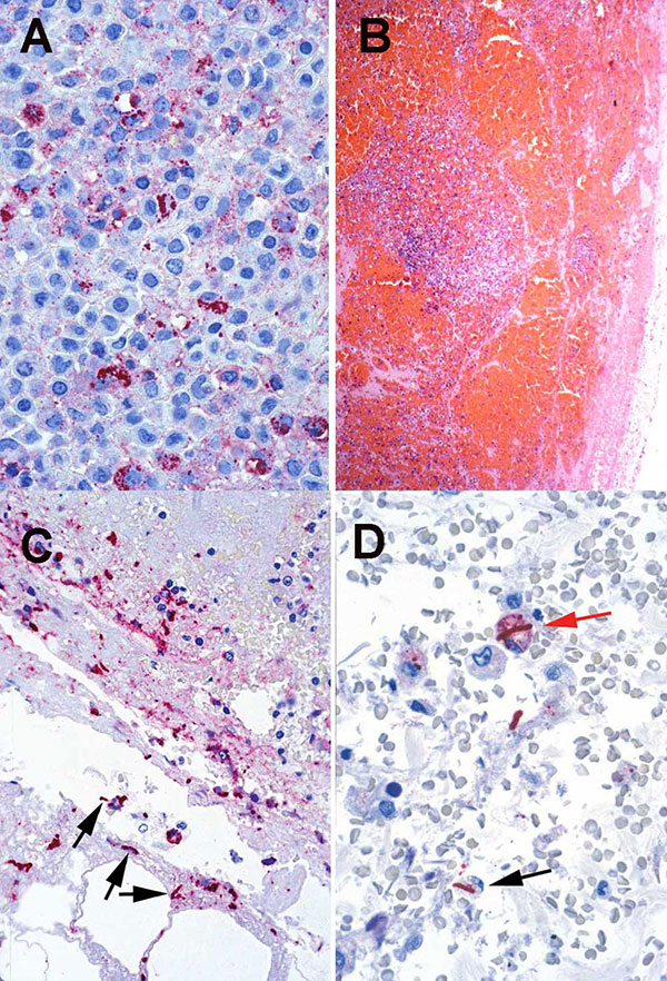
Figure 7. A. Pleural fluid cell block from a nonfatal case showing abundant Bacillus anthracis granular antigen staining inside mononuclear inflammatory cells. (Immunohistochemical assay with a mouse monoclonal anti-B. anthracis capsule antibody and detection with alkaline phosphatase and naphthol fast red, original magnification 158X). B. Mediastinal lymph node from a fatal case of anthrax showing extensive capsular and sinusoidal hemorrhage. (Hematoxylin and eosin, original magnification 25X). C. Lymph node from same case shown in B, showing abundant Bacillus anthracis granular antigen staining inside mononuclear inflammatory cells and bacilli (arrows) in the subcapsular hemorrhagic area. (Immunohistochemical assay with a mouse monoclonal anti-B. anthracis cell wall antibody and detection with alkaline phosphatase and naphthol fast red, original magnification 100X). D. Lung tissue from a fatal case showing Bacillus anthracis granular antigen staining inside a perihilar macrophage (red arrow) and intra- and extracellular bacilli (black arrow). (Immunohistochemical assay with a mouse monoclonal anti-B. anthracis cell-wall antibody and detection with alkaline phosphatase and naphthol fast red, original magnification 100X)
1Members of the team who contributed to the work presented in this manuscript are J. Aguilar, M. Andre, K. Baggett, B. Bell, D. Bell, M. Bowen, G. Carlone, M. Cetron, S. Chamany, B. De, C. Elie, M. Fischer, A. Hoffmaster, K. Glynn, R. Gorwitz, C. Greene, R. Hajjeh, T. Hilger, J. Kelly, R. Khabbaz, A. Khan, P. Kozarsky, M. Kuehnert, J. Lingappa, C. Marston, J. Nicholson, S. Ostroff, T. Parker, L. Petersen, R. Pinner, N. Rosenstein, A. Schuchat, V. Semenova, S. Steiner, F. Tenover, B. Tierney, T. Uyeki, S. Vong, D. Warnock, C. Spak, D. Jernigan, C. Friedman, M. Ripple, D. Patel, S. Pillai, S. Wiersma, R. Labinson, L. Kamal, E. Bresnitz, M. Layton, G. DiFerdinando, S. Kumar, P. Lurie, K. Nalluswami, L. Hathcock, L. Siegel, S. Adams, I. Walks, J. Davies-Coles, M. Richardson, K. Berry, E. Peterson, R. Stroube, H. Hochman, M. Pomeranz, A. Friedman-Kien, D. Frank, S. Bersoff-Matcha, J. Rosenthal, N. Fatteh, A. Gurtman, R. Brechner, C. Chiriboga, J. Eisold, G. Martin, K. Cahill, R. Fried, M. Grossman, and W. Borkowsky.